Alexandru V Avram1,2, Qiyuan Tian3, Qiuyun Fan3, Susie Y Huang3,4, and Peter J Basser1
1Eunice Kennedy Shriver National Institute of Child Health and Human Development, National Institutes of Health, Bethesda, MD, United States, 2Center for Neuroscience and Regenerative Medicine, The Henry Jackson Foundation, Bethesda, MD, United States, 3Athinoula A. Martinos Center for Biomedical Imaging, Department of Radiology, Massachusetts General Hospital, Harvard Medical School, Charlestown, MA, United States, 4Harvard-MIT Division of Health Sciences and Technology, Massachusetts Institute of Technology, Cambridge, MA, United States
1Eunice Kennedy Shriver National Institute of Child Health and Human Development, National Institutes of Health, Bethesda, MD, United States, 2Center for Neuroscience and Regenerative Medicine, The Henry Jackson Foundation, Bethesda, MD, United States, 3Athinoula A. Martinos Center for Biomedical Imaging, Department of Radiology, Massachusetts General Hospital, Harvard Medical School, Charlestown, MA, United States, 4Harvard-MIT Division of Health Sciences and Technology, Massachusetts Institute of Technology, Cambridge, MA, United States
We measure mean apparent diffusion
propagators (MAP) in the human brain using different diffusion times. We estimate
the time-scaling parameters of two important propagator parameters in order to
describe anomalous diffusion processes in fractal-like tissue environments.
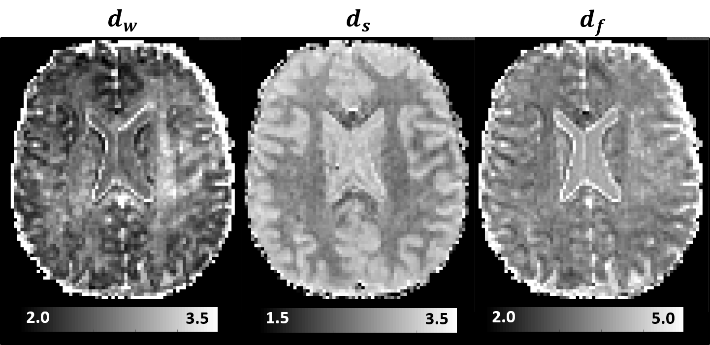
Figure 4: Time-scaling MRI parameters derived
from in vivo MAP-MRI data with
different diffusion times quantify anomalous diffusion in live brain tissues. The
random walk dimension, dw, and the spectral dimension, ds, are dynamic exponents that describe the diffusion process in a
fractal-like medium, while the fractal dimension, df, describes the scaling of the mass of the environment with
distance.
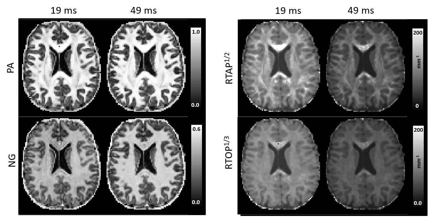
Figure 1: Diffusion time dependence of MAP-MRI scalar
parameters. Generally, the tissue contrast in Propagator Anisotropy (PA) and Non-Gaussianity (NG) images increases with diffusion time; meanwhile, both the Return-to-axis probability (RTAP) and the Return-to-origin-probability (RTOP) decrease with diffusion time throughout the parenchyma, especially in gray matter.