Francesco Grussu1, Kinga Bernatowicz1, Ignasi Barba2, and Raquel Perez-Lopez1,3
1Radiomics Group, Vall d'Hebron Institute of Oncology, Vall d'Hebron Barcelona Hospital Campus, Barcelona, Spain, 2NMR Lab, Vall d'Hebron Institute of Oncology, Vall d'Hebron Barcelona Hospital Campus, Barcelona, Spain, 3Department of Radiology, Hospital Universitari Vall d'Hebron, Barcelona, Spain
1Radiomics Group, Vall d'Hebron Institute of Oncology, Vall d'Hebron Barcelona Hospital Campus, Barcelona, Spain, 2NMR Lab, Vall d'Hebron Institute of Oncology, Vall d'Hebron Barcelona Hospital Campus, Barcelona, Spain, 3Department of Radiology, Hospital Universitari Vall d'Hebron, Barcelona, Spain
We investigate in simulations the feasibility of
estimating hepatocyte size and diffusivity from diffusion signal cumulants
(apparent diffusivity and kurtosis) at fixed diffusion time. The task appears to
be feasible with realistic liver SNR levels.
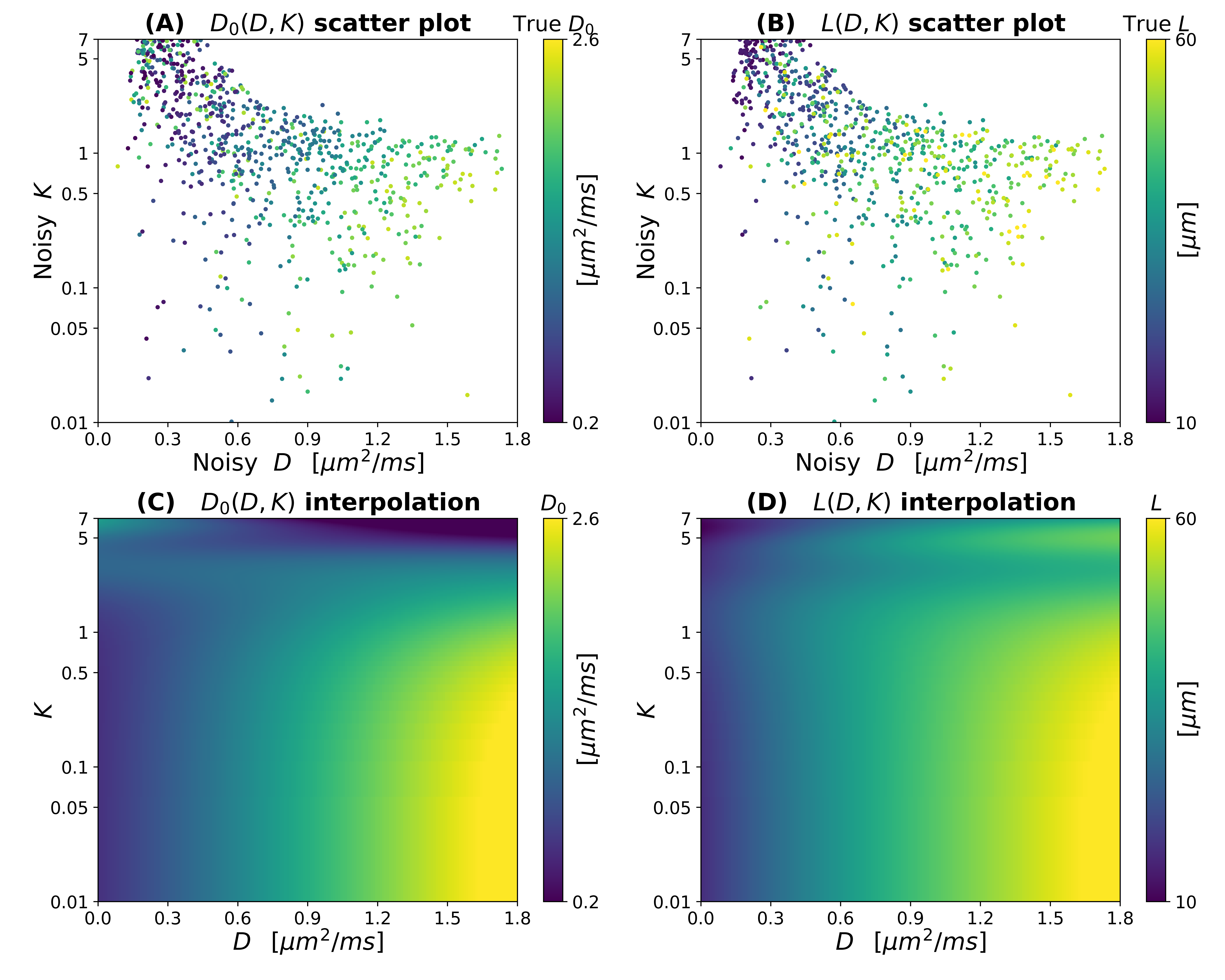
Figure
4: example of interpolation of the experimental D0(D,K) and L(D,K) observations for a SNR at b = 0 of 20.
Top: observations for Δ = 50 ms and bmax = 1500 s mm–2 (A, to the left: D0(D,K); B, to the right: L(D,K)). Bottom: radial basis
function interpolation and extrapolation to the whole (D,K) domain (C, to the left: D0(D,K); D, to the right: L(D,K)).
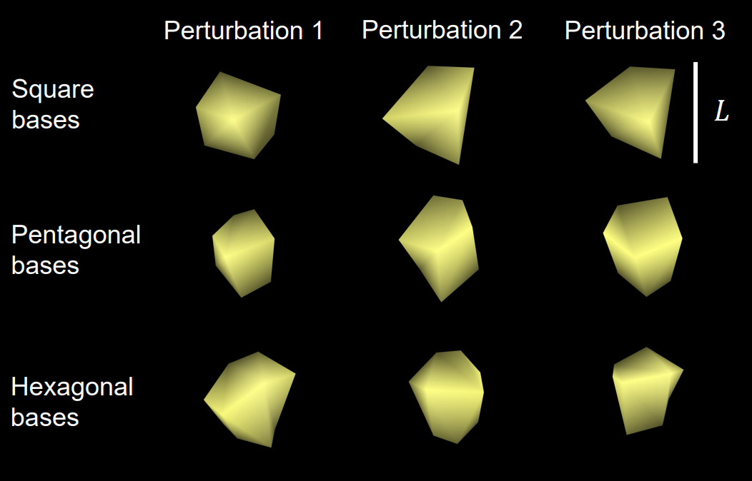
Figure
1: example of synthetic hepatic cells considered in this
study. From left to right: different cell realisations obtained by independent
perturbations of regular prisms with the same base shape. From top to bottom:
different base shapes (square, pentagonal and hexagonal). L corresponds to the inter-base distance.