Valerie Klein1,2, Mathias Davids1,2,3, Donald Straney2, Livia Vendramini2, Lothar R. Schad1, Maaike van den Boomen2,3,4, Christopher Nguyen2,3,4, Lawrence L. Wald2,3,5, and Bastien Guerin2,3
1Computer Assisted Clinical Medicine, Medical Faculty Mannheim, Heidelberg University, Mannheim, Germany, 2A. A. Martinos Center for Biomedical Imaging, Department of Radiology, Massachusetts General Hospital, Charlestown, MA, United States, 3Harvard Medical School, Boston, MA, United States, 4Cardiovascular Research Center, Cardiology Division, Massachusetts General Hospital, Charlestown, MA, United States, 5Harvard-MIT Division of Health Sciences and Technology, Cambridge, MA, United States
1Computer Assisted Clinical Medicine, Medical Faculty Mannheim, Heidelberg University, Mannheim, Germany, 2A. A. Martinos Center for Biomedical Imaging, Department of Radiology, Massachusetts General Hospital, Charlestown, MA, United States, 3Harvard Medical School, Boston, MA, United States, 4Cardiovascular Research Center, Cardiology Division, Massachusetts General Hospital, Charlestown, MA, United States, 5Harvard-MIT Division of Health Sciences and Technology, Cambridge, MA, United States
We developed a magnetic stimulator to measure
cardiac stimulation thresholds in pigs and to compare with simulations in
porcine-specific body and heart models created from MRI acquisitions.
Simulations of cardiac stimulation may help to inform appropriate safety limits
for MRI gradients.
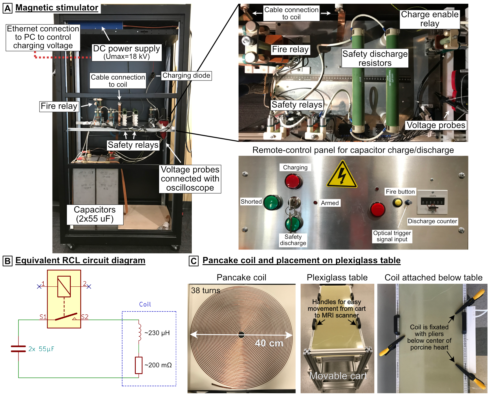
Figure 1: Magnetic
stimulator. A) The pulse generator consists of a capacitor bank (C=110 µF
total) that is charged with a high-voltage power supply. When the relay is
closed, the capacitors are discharged into a coil, creating a damped sinusoidal
coil current. The capacitor charge/discharge can be controlled remotely. B) RCL
circuit diagram for the capacitors, relay and the coil. C) Flat pancake
coil (Dout=40 cm, Din=2.5 cm, 38 turns of 2.3-mm copper
wire) and plexiglass table on which the pig is placed. The coil is attached
below the table and centered below the porcine heart.
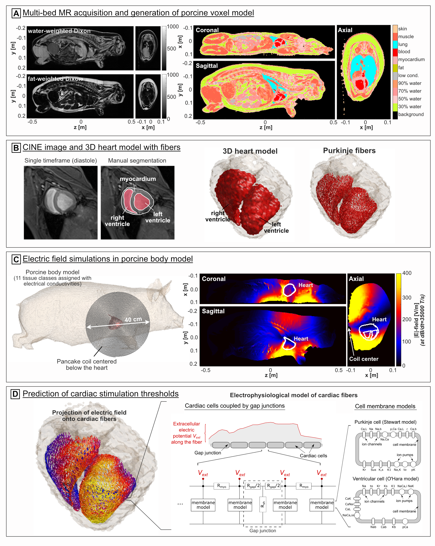
Figure 2: Simulation workflow. A) We acquired
multi-bed Dixon images to generate porcine voxel models including 11 different
tissue classes assigned with electrical conductivity values. B) We used
CINE images (diastolic phase) to create 3D heart models to which we added cardiac
Purkinje and ventricular muscle fibers networks. C) We simulated
electric fields induced in the porcine models by a 40-cm pancake coil. D) We
projected the electric fields onto the cardiac fibers and predict cardiac
stimulation thresholds using electrophysiological models of the cell membrane
ion dynamics.