Mingyan Li1, Ewald Weber1, Aurelien Destruel2, Feng Liu1, and Stuart Crozier1
1School of Information Technology and Electrical Engineering, The University of Queensland, Australia, Australia, 2Center for Magnetic Resonance in Biology and Medicine, Aix-Marseille University, MARSEILLE, France
1School of Information Technology and Electrical Engineering, The University of Queensland, Australia, Australia, 2Center for Magnetic Resonance in Biology and Medicine, Aix-Marseille University, MARSEILLE, France
Compared to supine position, the local SAR is raised when the patient is in standing position inside of the MRI-Linac coil, but is much lower than a standard 32-rung whole-body coil. SAR control strategy could be considered by using the vendor provided control measures for a standard coil.
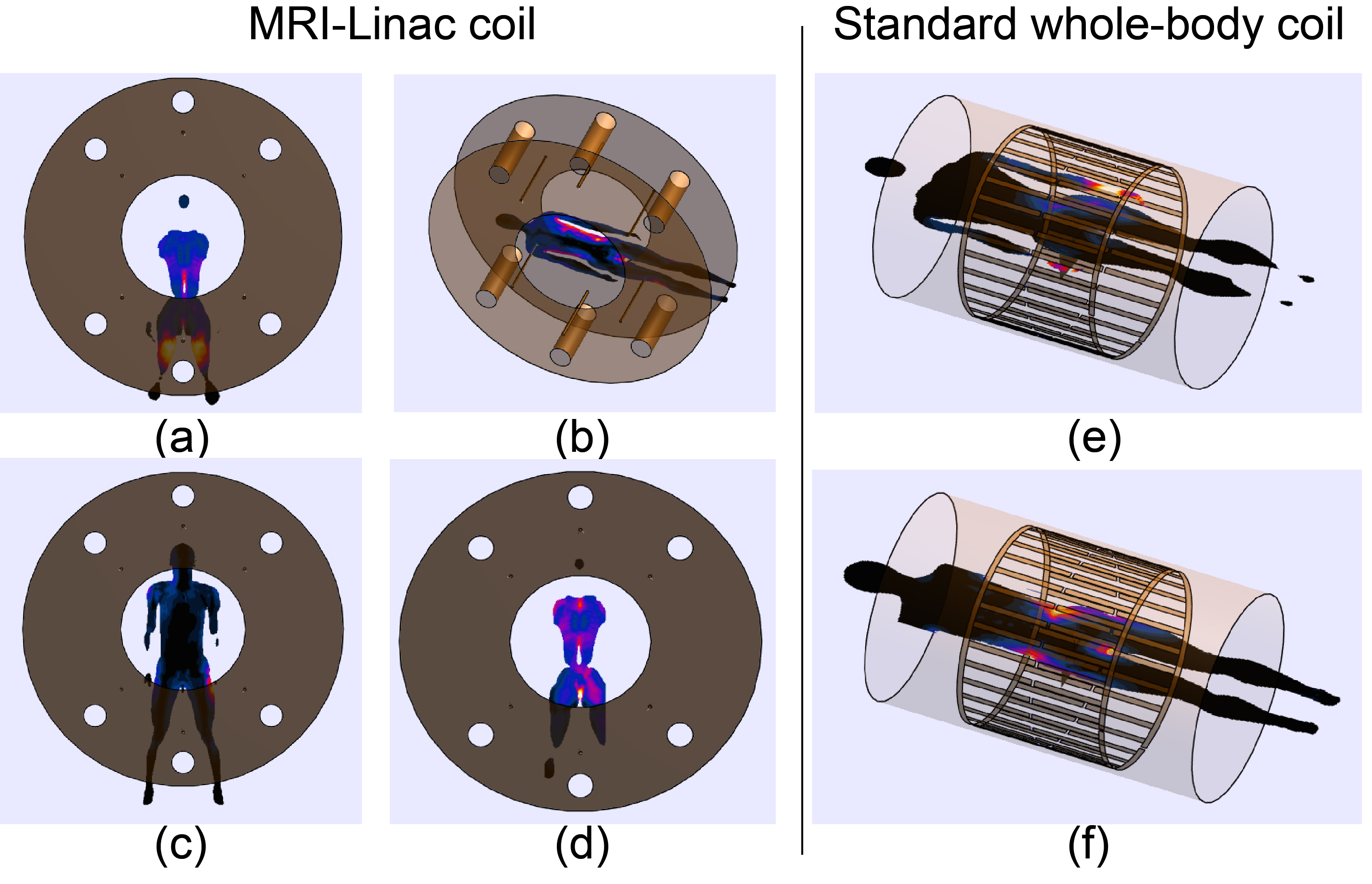
Figure 2 (a) SAR10g
distribution when d=15mm in standing position. (b) SAR10g
distribution in supine position. (c) SAR10g distribution when d =
225 mm in standing position. (d) SAR10g distribution (exclude hands)
when d = 225 mm in standing position. (e) SAR10g distribution with a
conventional whole-body coil in supine position. (f) SAR10g
distribution (exclude hands) with a conventional whole-body coil in supine
position. Legend is individually adjusted for best visualisation effect, the
explicit value can be found in Table I and II.
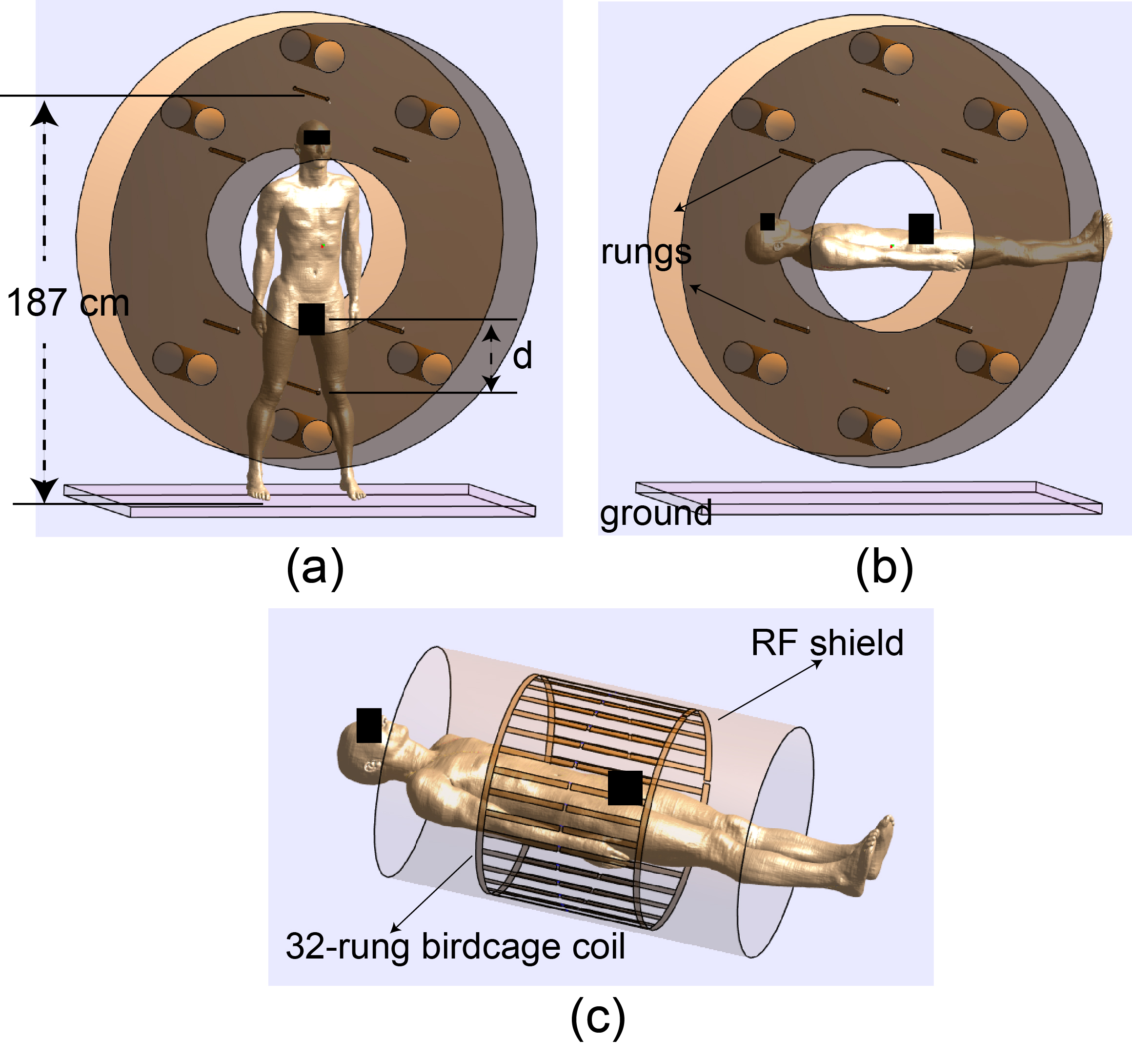
Figure
1. (a) Duke model in the MRI-Linac coil in the standing position. The distance
from the top rung to the ground is 187cm. The variable d denotes the distance
from the bottom rung to the genital area.
(b) Duke model in the MRI-Linac coil in supine position. (c) Duke model
in a standard 32-rung birdcage coil.