Tomohisa Okada1, Shinya Handa2, Bill Ding2, Shin-ichi Urayama1, Koji Fujimoto1, Atsushi Shima3, Takashi Ayaki4, Nobukatsu Sawamoto3, Ryosuke Takahashi4, Hirotaka Onoe1, Tadashi Isa1, and Labros Petropoulos2
1Human Brain Research Center, Kyoto University, Kyoto, Japan, 2Quality Electrodynamics, Mayfield Village, OH, United States, 3Department of Human Health Sciences, Kyoto University, Kyoto, Japan, 4Department of Neurology, Kyoto University, Kyoto, Japan
1Human Brain Research Center, Kyoto University, Kyoto, Japan, 2Quality Electrodynamics, Mayfield Village, OH, United States, 3Department of Human Health Sciences, Kyoto University, Kyoto, Japan, 4Department of Neurology, Kyoto University, Kyoto, Japan
Inductively coupled volume coils that can be inserted to the knee
coil was created. They could fine structures of the specimen using the same
sequence implemented on the whole-body human 7T scanner.
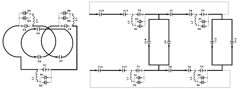
Figure 1. Designs of the coils
(left: 26 mm, right: 64 mm diameters; D26 and D64 coils, respectively).
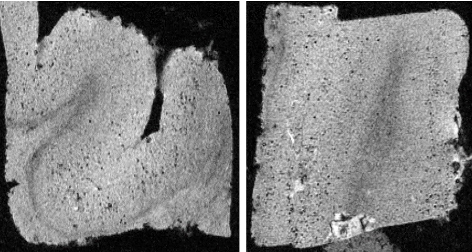
Figure
3. Brain specimen images of a
Alzheimer’s disease patient using the D26 coil in isotropic 50 μm
resolution (left: axial, right: coronal). Numerous tiny dots were
considered to be
iron-loaded amyloid plaques.