Yuto Murakami1 and Yasuhiko Terada1
1University of Tsukuba, Tsukuba, Japan
1University of Tsukuba, Tsukuba, Japan
We proposed a new double helix dipole coil
for high-field MR and constructed it for 7T system, which had nearly twice the
signal-to-noise ratio of a saddle coil. We demonstrated MR microimaging of
human embryo.
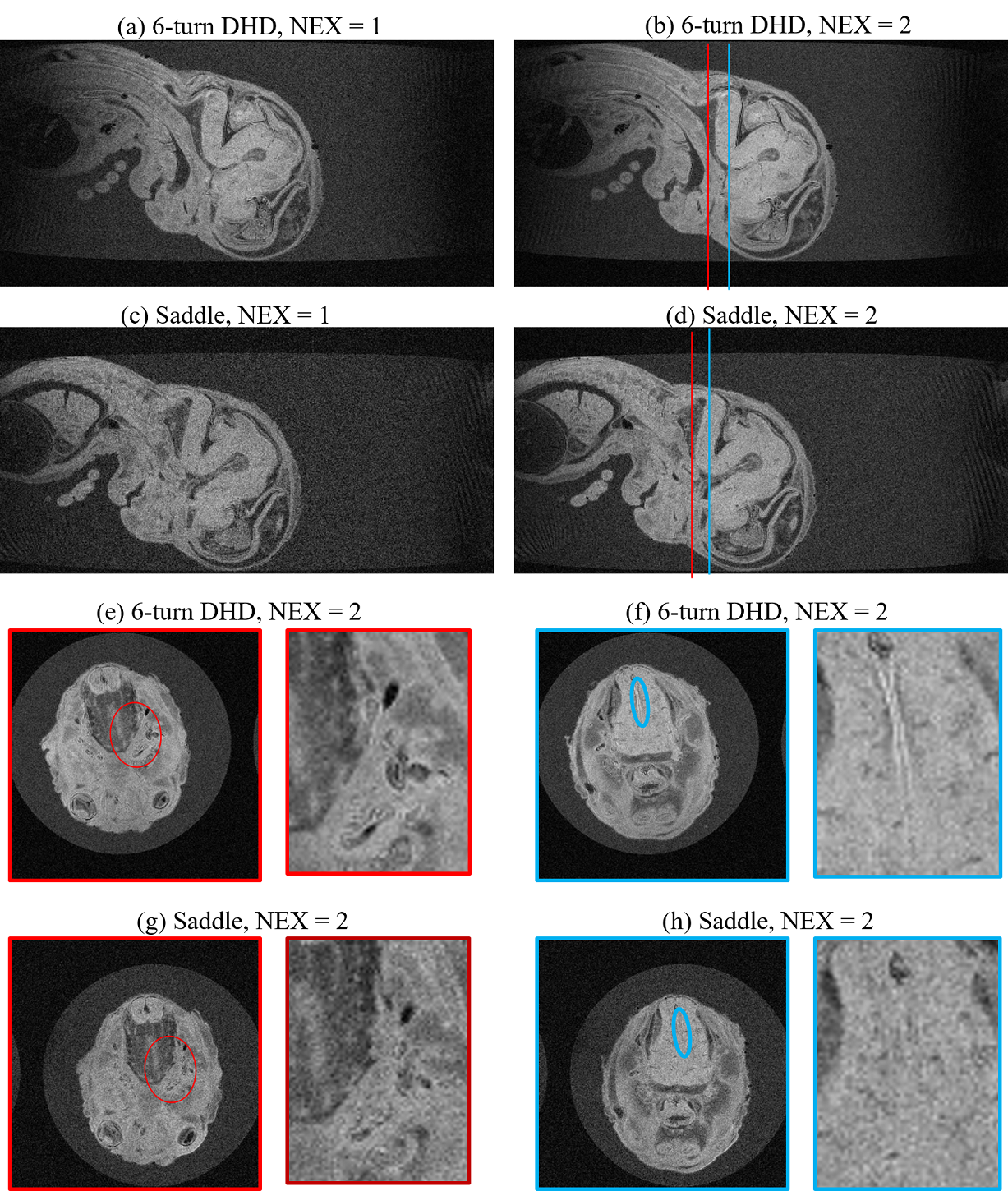
Fig. 5
MR microimaging results of the human embryo acquired with the 6-turn
and saddle coils. (a-d) Sagittal views. (e-h) Axial views. In Figs. (e)-(h),
the magnified images near the trigeminal fibers (red ovals) and the border of
the medulla oblongata (blue ovals) were shown on the right side of the original
images. NEX: number of excitations.
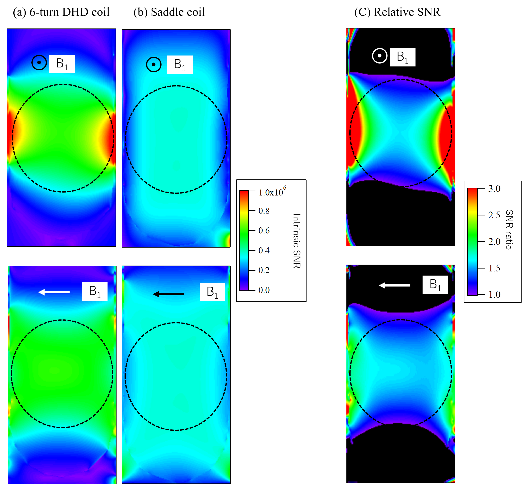
Fig. 3
Simulated SNR maps. (a,b) Intrinsic SNR maps for (a) 6-turn DHD and
(b) saddle coils. (c) Relative SNR of the 6-turn DHD coil with respect to the
saddle coil.