Daniel Papp1, Urs Schüffelgen2, Mo Shahdloo2, Sebastian W Rieger1,3, Aaron T Hess4, Matthew Rushworth2, and Stuart Clare1
1Wellcome Centre for Integrative Neuroimaging, FMRIB, Nuffield Department of Clinical Neurosciences, University of Oxford, Oxford, United Kingdom, 2Wellcome Centre for Integrative Neuroimaging, Department of Experimental Psychology, University of Oxford, Oxford, United Kingdom, 3Wellcome Centre for Integrative Neuroimaging, OHBA, Department of Psychiatry, University of Oxford, Oxford, United Kingdom, 4British Heart Foundation Centre of Research Excellence, Department of Cardiovascular Medicine, University of Oxford, Oxford, United Kingdom
1Wellcome Centre for Integrative Neuroimaging, FMRIB, Nuffield Department of Clinical Neurosciences, University of Oxford, Oxford, United Kingdom, 2Wellcome Centre for Integrative Neuroimaging, Department of Experimental Psychology, University of Oxford, Oxford, United Kingdom, 3Wellcome Centre for Integrative Neuroimaging, OHBA, Department of Psychiatry, University of Oxford, Oxford, United Kingdom, 4British Heart Foundation Centre of Research Excellence, Department of Cardiovascular Medicine, University of Oxford, Oxford, United Kingdom
We present the evaluation of a 15ch macaque head coil for awake behaving fMRI. The coil shows marked improvement over our current hardware, and offers highly accelerated imaging capability, up to a combined factor of Multiband 6 and in-plane GRAPPA 2
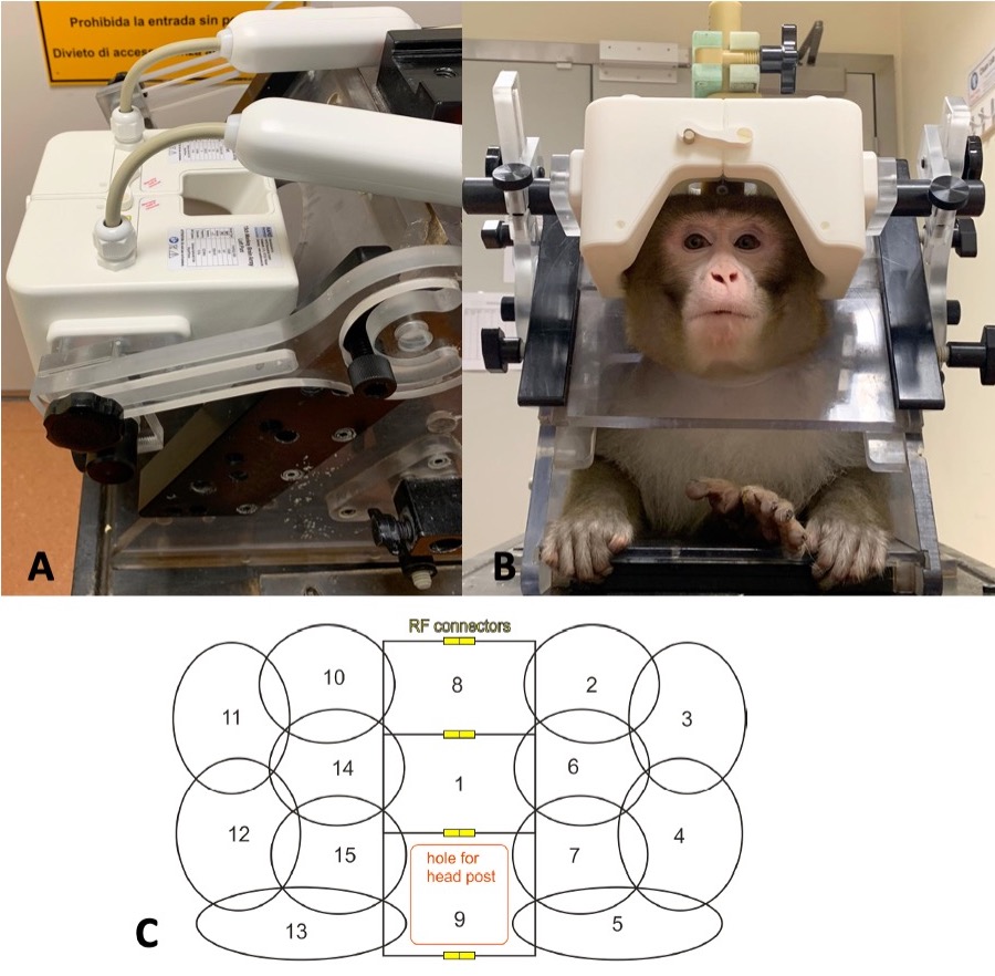
Figure 1: The fit of the bespoke coil and the animal chair (A), a macaque in the coil (B), and a schematic showing the receiver loops (C)
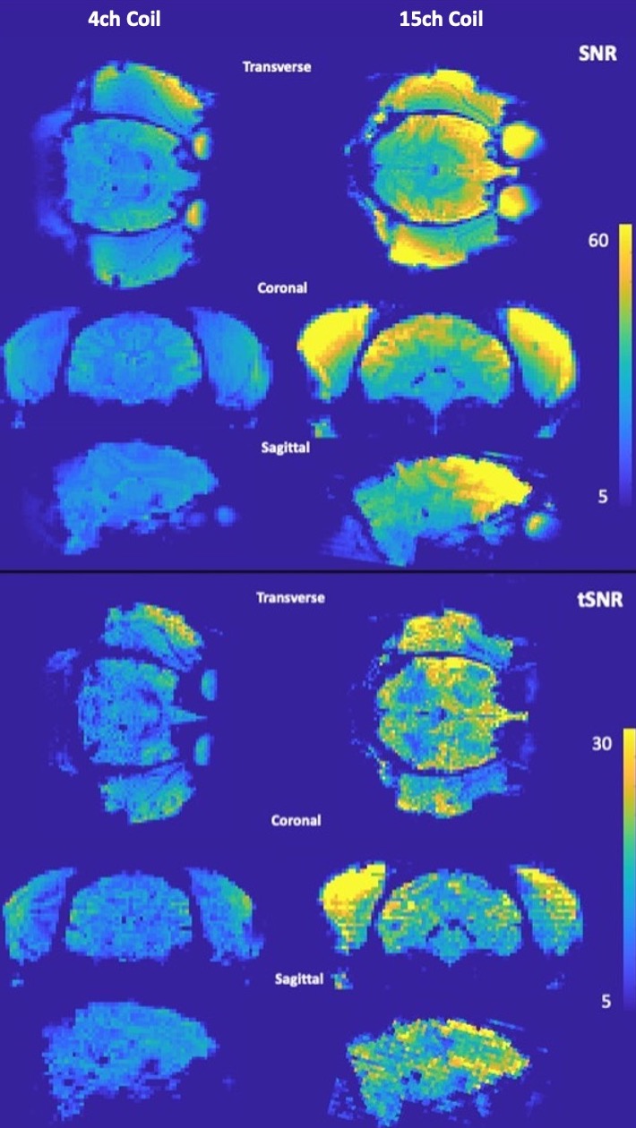
Figure 3: Transverse, coronal and sagittal views of the SNR (first three rows) and tSNR (last three rows) maps of the head for in-vivo scans acquired using the 4-channel flexible coil (first column) and the 15-channel coil (second column). The improvements are most pronounced in the frontal regions, however, a meaningful increase in imaging performance can be observed through the brain. Note that stock reconstruction was used, and the images were not brain extracted.