Kanishka Sharma1, Bashair Alhummiany2, David Shelley2,3, Margaret Saysell2,3, Maria-Alexandra Olaru4, Bernd Kühn4, Julie Bailey3, Kelly Wroe3, Cherry Coupland3, Michael Mansfield3, and Steven Sourbron1
1Department of Imaging, Infection, Immunity and Cardiovascular Disease, University of Sheffield, Sheffield, United Kingdom, 2Department of Biomedical Imaging Sciences, University of Leeds, Leeds, United Kingdom, 3Leeds Teaching Hospitals, Leeds, United Kingdom, 4Siemens Healthcare GmbH, Erlangen, Germany
1Department of Imaging, Infection, Immunity and Cardiovascular Disease, University of Sheffield, Sheffield, United Kingdom, 2Department of Biomedical Imaging Sciences, University of Leeds, Leeds, United Kingdom, 3Leeds Teaching Hospitals, Leeds, United Kingdom, 4Siemens Healthcare GmbH, Erlangen, Germany
The results indicate overall
comparable repeatability for MRI biomarkers of renal tissue structure and perfusion
using phase contrast, while also highlighting the need for formal MRI quality
assurance prior to image processing.
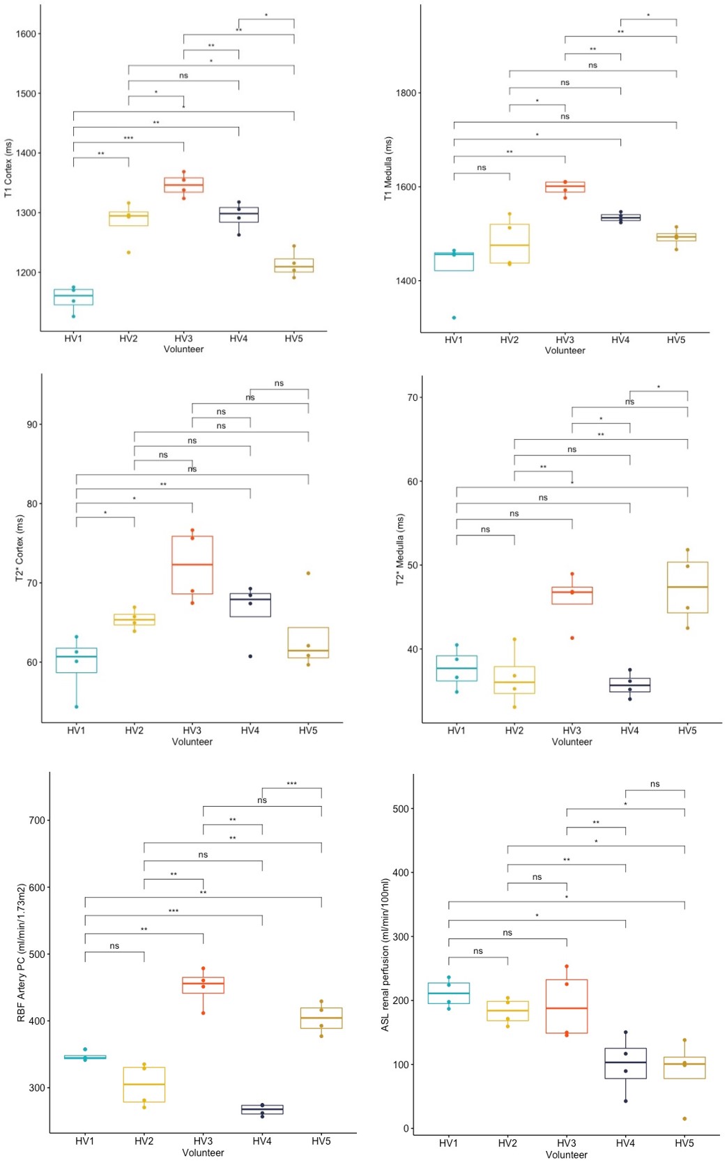
Figure 3. Box plots for T1, T2*
mapping in the renal cortex and medulla (ROIs), arterial RBF (BSA normalised)
using PC-MRI, and renal perfusion (ml/min/100ml) with ASL, from 4 repeatability
measurements in 5 healthy volunteers (HV) on the reference MRI scanner (MAGNETOM Prisma 3T, Siemens
Healthcare GmbH, Erlangen, Germany) using the iBEAt MRI protocol. Pairwise comparison using
t-test shows the statistical significance of differences (ns: not significant =
p > 0.05; *p ≤ 0.05; **p ≤ 0.01; ***p ≤ 0.001) between HVs.
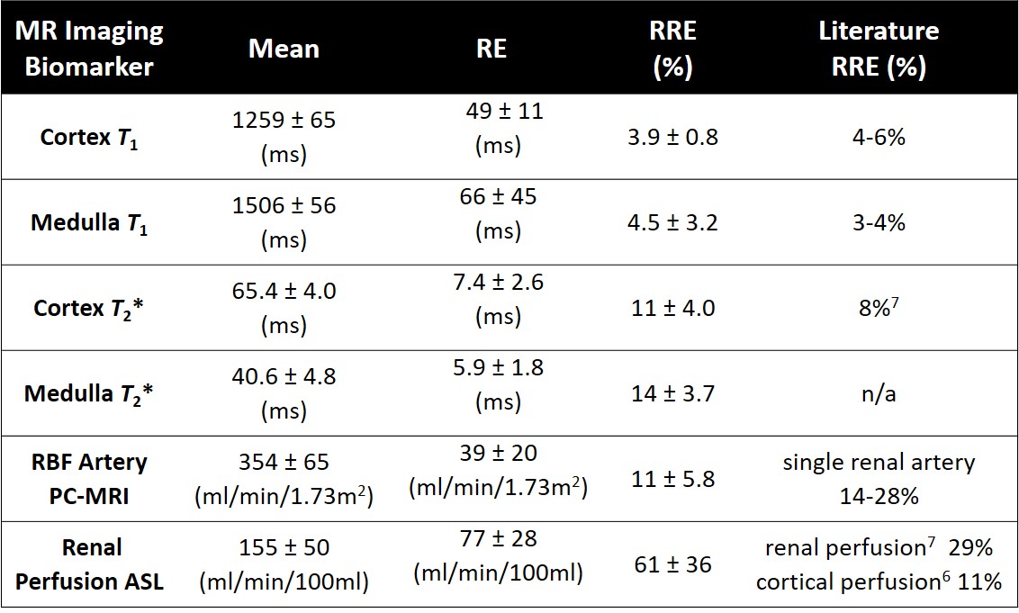
Table 1. 95% confidence interval for the mean value of each
parameter (1st column), and for the mean value of their RE (2nd
column) and RRE (3rd column). Literature values of RRE (4th column).