Pete Thelwall1,2, Jehill Parikh1, Benjamin Pippard1, Caroline Wroe3, Rob Janiczek4, Steven Sourbron5, and Neil Sheerin1
1Translational and Clinical Research Institute, Newcastle University, Newcastle upon Tyne, United Kingdom, 2Centre for In Vivo Imaging, Newcastle University, Newcastle upon Tyne, United Kingdom, 3South Tees Hospitals NHS Foundation Trust, Middlesborough, United Kingdom, 4GlaxoSmithKline, Philadelphia, PA, United States, 5University of Sheffield, Sheffield, United Kingdom
1Translational and Clinical Research Institute, Newcastle University, Newcastle upon Tyne, United Kingdom, 2Centre for In Vivo Imaging, Newcastle University, Newcastle upon Tyne, United Kingdom, 3South Tees Hospitals NHS Foundation Trust, Middlesborough, United Kingdom, 4GlaxoSmithKline, Philadelphia, PA, United States, 5University of Sheffield, Sheffield, United Kingdom
A multiparametric kidney scan protocol was implemented. An increase in cortical native T1 and T2 was observed in patients with moderate renal impairment and biopsy-proven fibrosis compared to healthy controls.
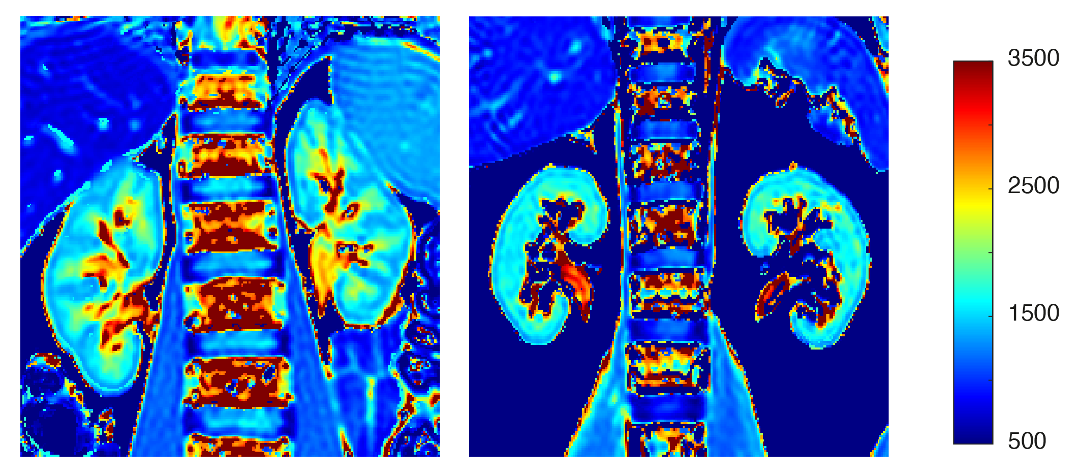
T1 maps from a healthy volunteer (left) and patient with moderate renal impairment and biopsy-proven fibrosis (right). Lower contrast (ie. smaller T1 different) between cortical and medullary regions is observed in the patient with renal disease compared to the healthy volunteer.
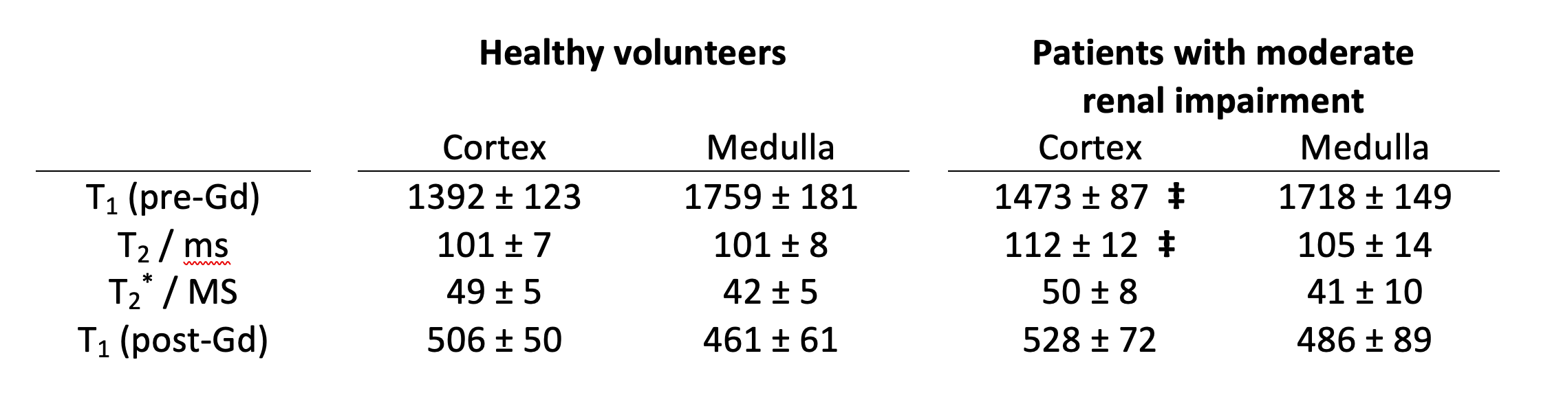
Cortical and medullary T1 (pre- and post-administration of gadolinium-based contrast agent), T2 and T2* measurements from healthy volunteers and patients with moderate renal impairment. ‡ denotes a statistically significant different from healthy controls (p < 0.05).