Haonan Zhang1, Qingwei Song1, Jiazheng Wang2, Zhiwei Shen2, Renwang Pu1, Nan Zhang1, and Ailian Liu1
1Department of Radiology, the First Affiliated Hospital of Dalian Medical University, Dalian, China, 2PHILIPS——Philips Healthcare, beijing, China
1Department of Radiology, the First Affiliated Hospital of Dalian Medical University, Dalian, China, 2PHILIPS——Philips Healthcare, beijing, China
In
this study, we evaluated the image quality of renal artery based on B-TFE sequence with SENSE and CS-SENSE.
CS factor of 6 is recommended for clinical renal artery imaging based on B-TFE
sequence.
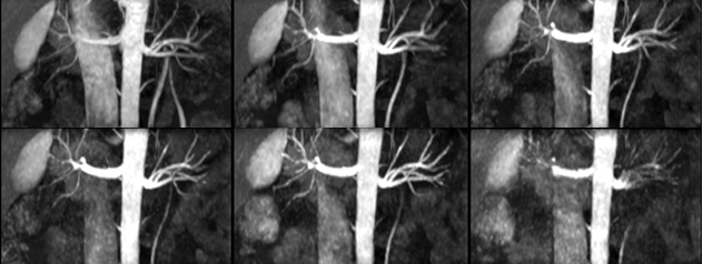
Figure2. Reconstruction
of renal artery. The first row,
from left to right: SENSE2, CS2-CS4. The second row, from left to right: CS6-CS10.
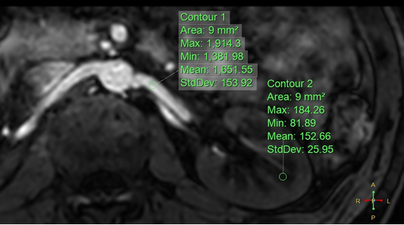
Figure 1.
Location of two ROI of the blood
vessels and the renal medulla. Use ROI to measure SI and SD on the left
sides. The measured left blood vessel SI value was 1651.55, muscle SI value was
152.66, and muscle SD value was 25.95.