Nathan Sennesael1,2, Pieter R.E. De Backer3,4,5, Charlotte Debbaut4,6, Karel Decaestecker3,5, Pieter L.J. De Visschere2,7, Marijn M. Speeckaert8,9, Saar Vermijs4,5, Geert Villeirs2,7, and Pim Pullens1,2,7
1GIfMI, Ghent University, Ghent, Belgium, 2Diagnostic Sciences, Ghent University, Ghent, Belgium, 3Urology, Ghent University Hospital, Ghent, Belgium, 4IBiTech-bioMMeda, Ghent University, Ghent, Belgium, 5Human Structure and Repair, Ghent University, Ghent, Belgium, 6Cancer Research Institute Ghent (CRIG), Ghent University, Ghent, Belgium, 7Radiology, Ghent University Hospital, Ghent, Belgium, 8Nephrology, Ghent University Hospital, Ghent, Belgium, 9Internal diseases and Paediatrics, Ghent University, Ghent, Belgium
1GIfMI, Ghent University, Ghent, Belgium, 2Diagnostic Sciences, Ghent University, Ghent, Belgium, 3Urology, Ghent University Hospital, Ghent, Belgium, 4IBiTech-bioMMeda, Ghent University, Ghent, Belgium, 5Human Structure and Repair, Ghent University, Ghent, Belgium, 6Cancer Research Institute Ghent (CRIG), Ghent University, Ghent, Belgium, 7Radiology, Ghent University Hospital, Ghent, Belgium, 8Nephrology, Ghent University Hospital, Ghent, Belgium, 9Internal diseases and Paediatrics, Ghent University, Ghent, Belgium
We found that high resolution 3D-T2w-MRI scans in combination with artery-selective contrast injections is an effective alternative to ex-vivo kidney casting as it allows for 3D mapping of the renal vascular dominant regions and shows potential for segmentation of the renal vascular tree.
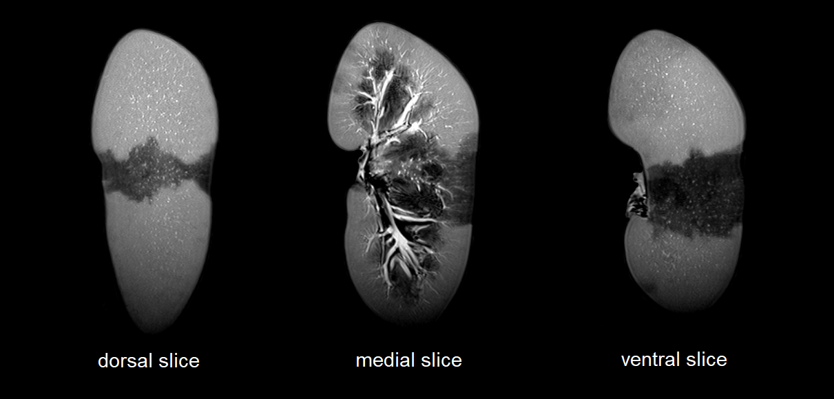
Figure 2: Frontal views of scan in which upper and lower segmental arteries are injected with water.
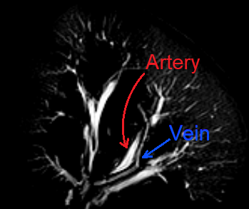
Figure 4: Visualization of the renal vascular tree