Ernest Eng1, Raimo Salo1, Heikki Tanila1, Mikko Kettunen1, and Olli Gröhn1
1A.I. Virtanen Institute for Molecular Sciences, Finland, Kuopio, Finland
1A.I. Virtanen Institute for Molecular Sciences, Finland, Kuopio, Finland
Knowledge
about how tau affects the microstructure is limited. Our anatomical
and diffusion data suggests that tauopathy results in structural and microstructural changes in the hTau.P301S-Tg mouse model’s inter-cerebellar
fibres, and possibly associates with the model’s motor declines.
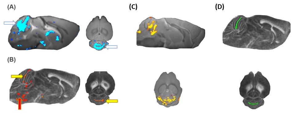
Fig 1. Group level pixel-wise analyses of
differences between controls and hTau.P301S mice at 5 mths of age as
exhibited by (A) Jacobian determinants showing local volumetric differences
around inter-cerebellar fibres (white arrows), (B) FA maps depicting microstructural
differences in inter-cerebellar fibres (yellow arrows) and brain stem (red
arrow), (C) local volumetric changes as depicted by Jacobian determinants from
2.5 – 5 mths, (D) location of Region of Interest (ROI) analysed; in (A), (B)
and (C) all coloured pixels statistically significant p<0.05, TFCE-corrected
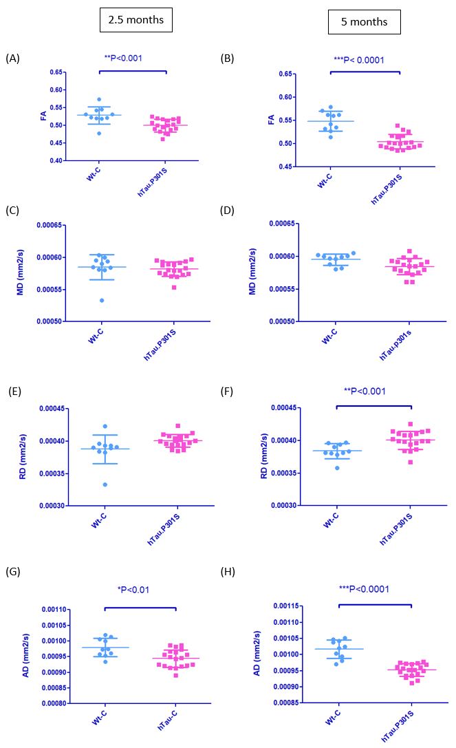
Fig 2. ROI analyses of DTI metrices in
inter-cerebellar fibres in controls and hTau.P301S mice. ROI is defined in
Fig 1D. (A) Fractional Anisotropy (FA) values at 2.5 mths, (B) FA values at 5
mths of age, (C) Mean Diffusivity (MD) values at 2.5 mths, (D) MD values at 5
mths of age, (E) Radial Diffusivity (RD) values at 2.5 mths, (F) RD values at 5
mths of age, (G) Average Diffusivity (AD) values at 2.5 mths, and (H) AD values
at 5 mths of age, data statistically significant at p<0.05, FDR-corrected