Jean-Baptiste Pérot1, Marina Célestine1, Miriam Riquelme-Pérez1, Carole Escartin1, Marc Dhenain1, Emmanuel Brouillet1, and Julien Flament1
1Université Paris-Saclay, Commissariat à l’Energie Atomique et aux Energies Alternatives (CEA), Centre National de la Recherche Scientifique (CNRS), Molecular Imaging Research Center (MIRCen), Laboratoire des Maladies Neurodégénératives, Fontenay-aux-Roses, France
1Université Paris-Saclay, Commissariat à l’Energie Atomique et aux Energies Alternatives (CEA), Centre National de la Recherche Scientifique (CNRS), Molecular Imaging Research Center (MIRCen), Laboratoire des Maladies Neurodégénératives, Fontenay-aux-Roses, France
Our imaging protocol combining rs-fMRI and DTI in a mouse model of Huntington's Disease (HD) evidenced vulnerable brain networks. FC loss and RD modifications in the white matter of zQ175 point out the key role of brain connectivity in HD.
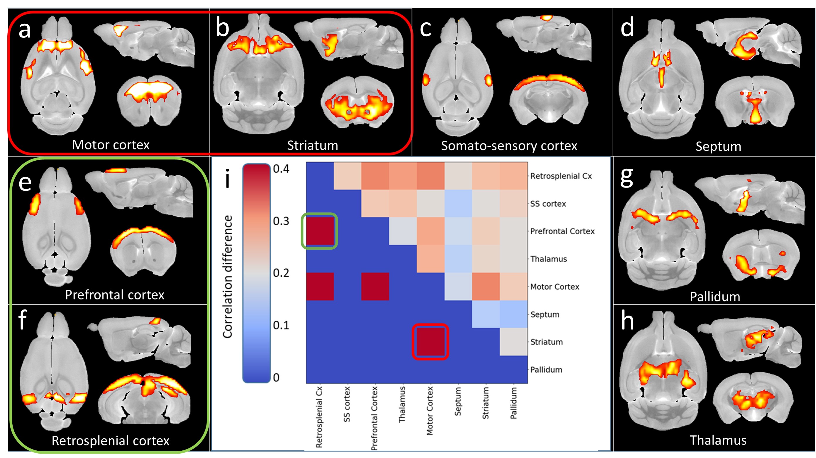
Figure 2: Dictionary Learning analysis of rs-fMRI data. a-h)
Regions extracted from 10 components DL. i) top-right: Functional connectivity
of extracted regions’ difference between WT and zQ175. Bottom-left:
representation of significant (red) and non-significant (blue) differences. Red
and green frames highlight somato-motor and Default Mode networks respectively.
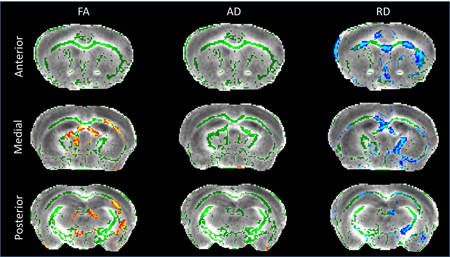
Figure 3:
Diffusion Tensor Imaging results after TBSS analysis. Green regions represent mean white matter skeleton as used in TBSS pipeline. Red-yellow zones show clusters of
voxels with significant decrease of FA in zQ175 mice. Blue-light blue zones show clusters of
voxels with increased RD in zQ175 mice (p<0.05).