Tomokazu Tsurugizawa1, Yuki Nakamura2,3,4, Yukari Nakamura2,3,4, Assunta Pelosi2,3,4, Boucif Djemai5, Clement Debacker6, Jean-Antoine Girault2,3,4, and Denis Herve2,4,7
1Human Informatics and Interaction Research Institute, National Institute of Advanced Industrial Science and Technology (AIST), Tsukuba, Japan, 2Inserm UMR-S 1270, Paris, France, 3Sciences and Technology Faculty, Sorbonne Universite, Paris, France, 4Institut du Fer à Moulin, Paris, France, 5NeuroSpin/CEA-Saclay, Gif-sur-Yvette, France, 6Inserm, UMR1266, Paris, France, 7Sorbonne Universite, Paris, France
1Human Informatics and Interaction Research Institute, National Institute of Advanced Industrial Science and Technology (AIST), Tsukuba, Japan, 2Inserm UMR-S 1270, Paris, France, 3Sciences and Technology Faculty, Sorbonne Universite, Paris, France, 4Institut du Fer à Moulin, Paris, France, 5NeuroSpin/CEA-Saclay, Gif-sur-Yvette, France, 6Inserm, UMR1266, Paris, France, 7Sorbonne Universite, Paris, France
Ipsilateral thalamic
nuclei are key regions for functional and structural alterations in basal
ganglia-thalamo-cortical loop in hemiparkinsonian mice.
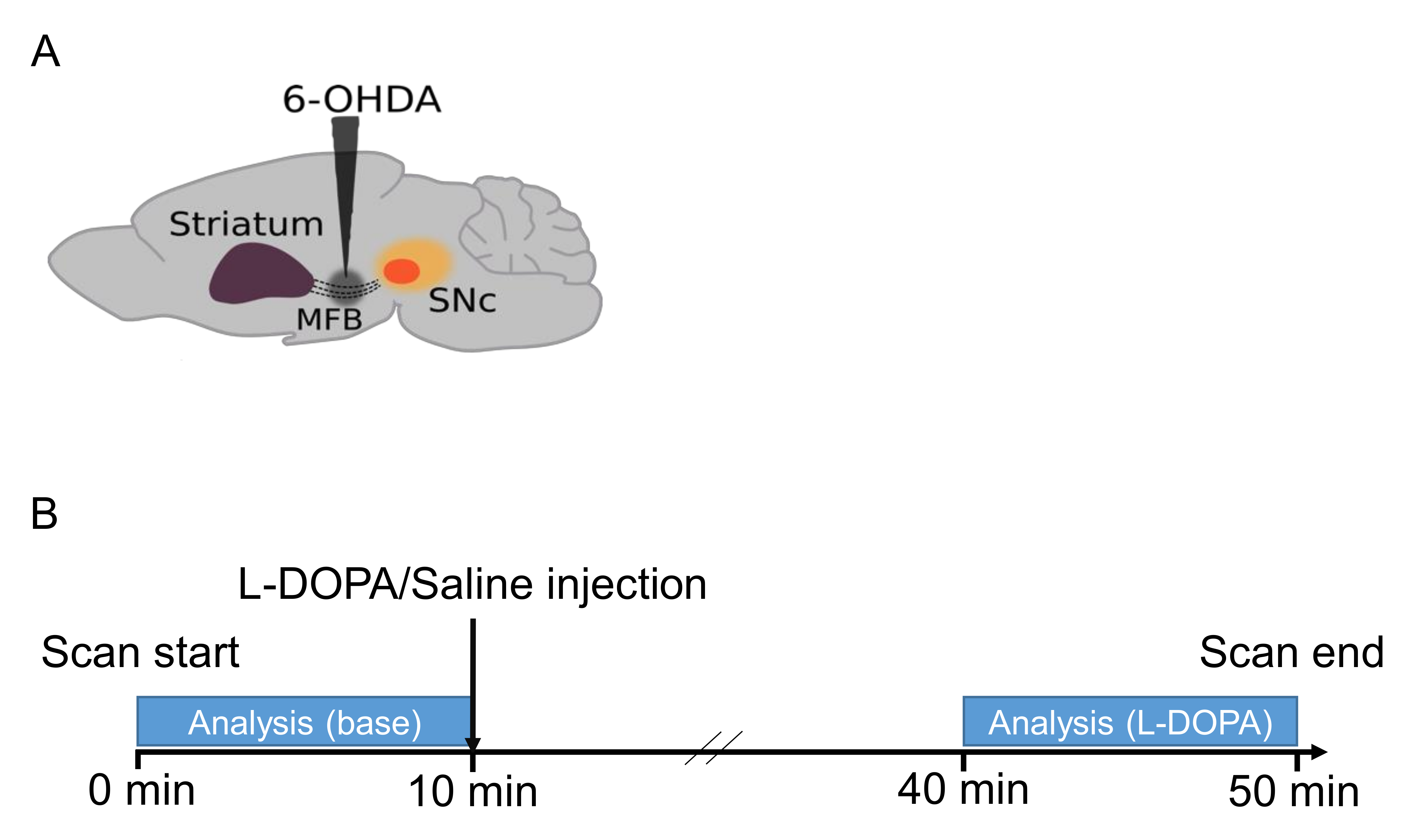
Figure
1
(A) 6-OHDA is microinjected into
the medial forebrain bundle (MFB) to unilaterally lesion dopamine ascending
pathways. SNc, substantia nigra pars compacta. (B) Schema of experimental
protocol. fMRI data were acquired for 50 min. For functional connectivity analysis, BOLD images were analyzed in
two time-windows of 10 min, 10 min before and 30 min after a L-DOPA injection.
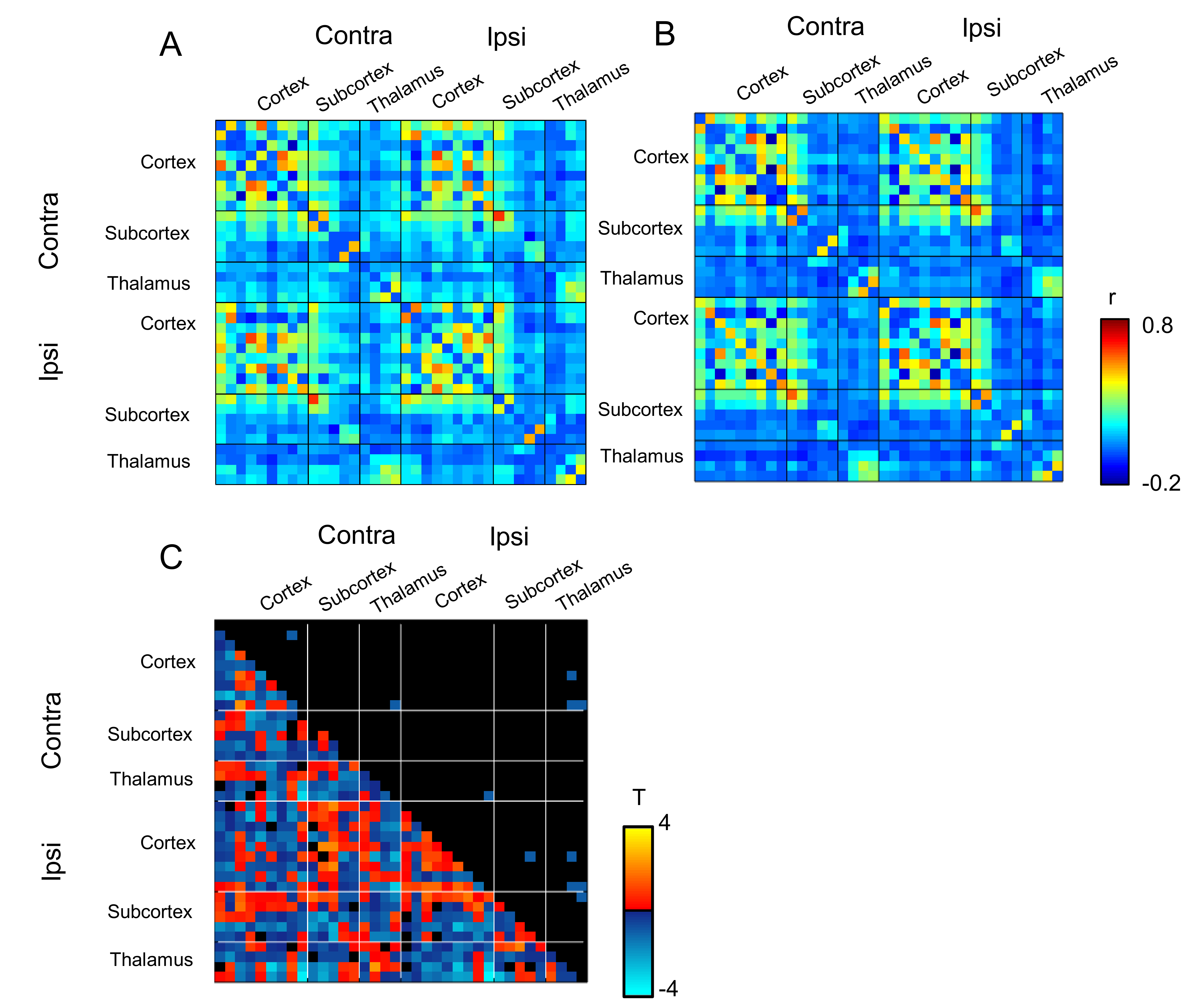
Figure
3
ROI-ROI matrices of correlation
coefficients in (A) sham-operated mice and (B) 6-OHDA-lesioned mice before
injection. Color bar, correlation coefficient. Thirty-six ROIs were classified
in the cortex (including somatosensory
cortex), subcortex (including striatum, GPi, GPe, SNR and SNC), and thalamus
(including STN, MD and CM). (C) Differences between 6-OHDA-lesioned and sham
mice before L-DOPA treatment. Left lower triangle panel shows t-values and
right upper triangle panel shows significant difference (p < 0.05, network
based statistic). Color bar, t-values.