Nicholas Vidas-Guscic1, Johan Van Audekerke1, Ben Jeurissen2, Jasmien Orije1, Tamara Vasilkovska1, Dorian Pustina3, Haiying Tang3, Roger Cachope3, Longbin Liu3, Mette Skinbjerg3, Celia Dominguez3, Ignacio Munoz-Sanjuan3, Annemie Van der Linden1, and Marleen Verhoye1
1Bio-Imaging Lab, university of Antwerp, Antwerp, Belgium, 2Vision Lab, university of Antwerp, Antwerp, Belgium, 3CHDI foundation, Princeton, NJ, United States
1Bio-Imaging Lab, university of Antwerp, Antwerp, Belgium, 2Vision Lab, university of Antwerp, Antwerp, Belgium, 3CHDI foundation, Princeton, NJ, United States
Whole-brain and region-based diffusion tensor,
diffusion kurtosis and fixel-based analysis indicate the presence of micro- and
macrostructural differences in many fiber populations
throughout the brain in the zQ175 mouse model of Huntington's disease.
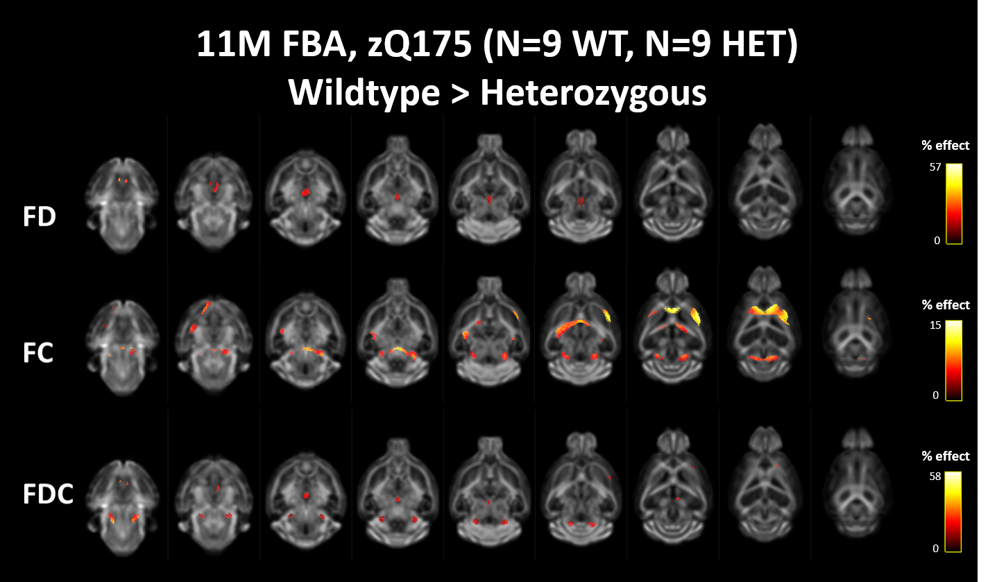
Figure 2: Fixel-based
analysis outcome for comparison zQ175 WT>HET. The
statistical maps show significant differences (wildtype > heterozygous) for
fiber density (FD) (top), fiber cross-section (FC) (middle) and the (fiber
density x cross-section) (FDC) combination metric (bottom). Significant fixels
(PFWE<0.05) are mapped to a 200.000 streamline tractogram and
coloured based on their percentage effect and superimposed on a fiber-orientation
distribution (fod) template (n=9 WT, n=9 HET).
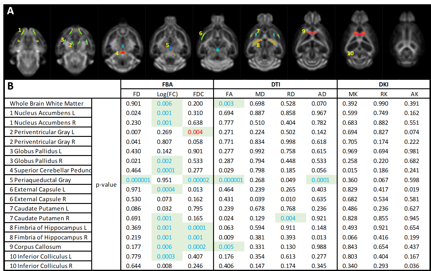
Figure 3: Region based
analysis of fixel, tensor and kurtosis metrics. A) Overview
of tracts of interest, selected following FBA. B) P-values corresponding to
unpaired t-tests (wildtype vs heterozygous) for each metric in each
region/tract of interest are displayed in the table. Green boxes indicate the
statistical tests that survive FDR correction. Blue and red characters indicate
a resp. lower or higher group average in zQ175 heterozygous compared to zQ175
wildtype animals.