Yuning Gu1, Lulu Wang2, Hongyi Yang2, Yun Wu2, Yong Chen3, Kai Zhong2, and Xin Yu1,3,4
1Biomedical Engineering, Case Western Reserve University, Cleveland, OH, United States, 2High Magnetic Field Laboratory, Chinese Academy of Sciences, Hefei, China, 3Radiology, Case Western Reserve University, Cleveland, OH, United States, 4Physiology and Biophysics, Case Western Reserve University, Cleveland, OH, United States
1Biomedical Engineering, Case Western Reserve University, Cleveland, OH, United States, 2High Magnetic Field Laboratory, Chinese Academy of Sciences, Hefei, China, 3Radiology, Case Western Reserve University, Cleveland, OH, United States, 4Physiology and Biophysics, Case Western Reserve University, Cleveland, OH, United States
This
study developed a 3D MRF method for T1 and T2 mapping of
monkey brain at 9.4 T. The high SNR
provided by a conformal head coil enabled whole-brain coverage at 0.35x0.35x1
mm3 resolution.
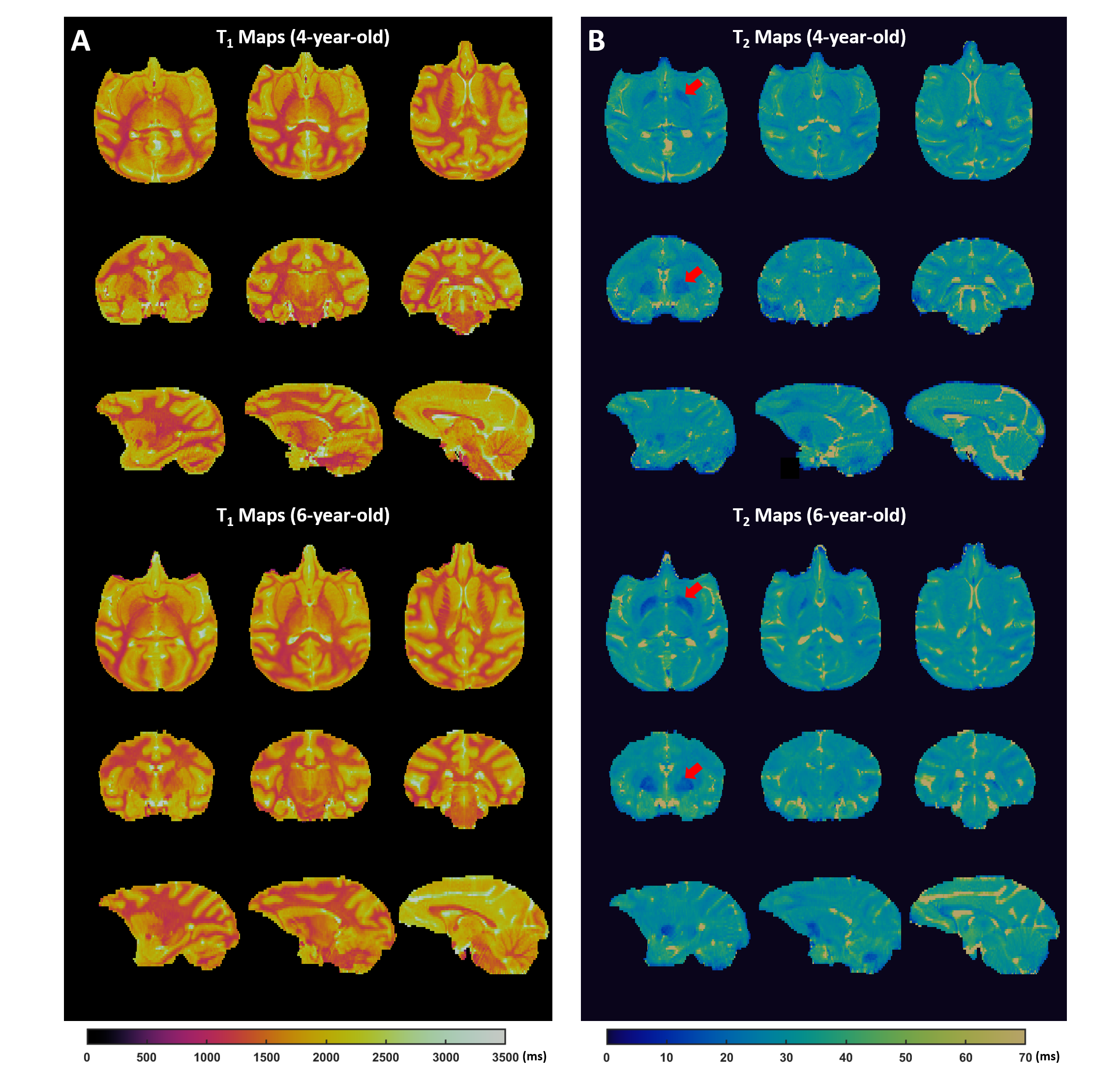
Figure 4. In vivo results. A&B. T1 (A) and T2 (B) maps of a 4- (top) and 6-year-old (bottom) monkey in the coronal, axial, and sagittal views. The red arrow indicates lower T2 value in globus pallidus in the 6-year-old monkey.
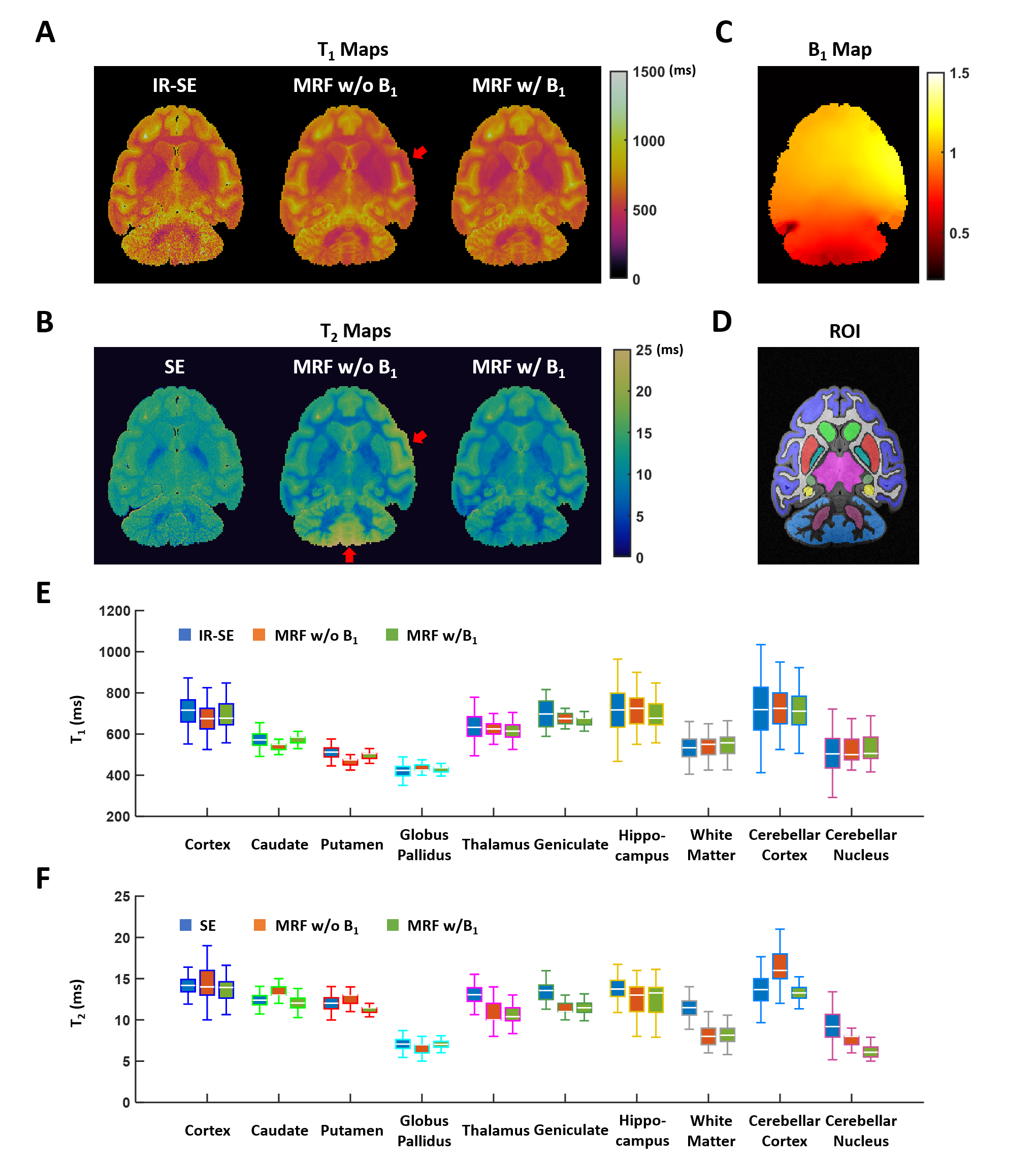
Figure 3. Ex vivo results. A&B. T1 (A) and T2 (B) maps by IR-SE (T1) or SE (T2), MRF without B1 correction, and MRF with B1 correction. The red arrow indicates T2 overestimation. C. Maps of B1 factor of the conformal coil. D. Color-coded ROIs. E&F. Boxplots of T1 (E) and T2 (F) values in selected ROIs.