Patrick McCunn1, Xiaoyun Xu2, Alex Li2, Arthur Brown2, and Robert Bartha2
1Patrick McCunn, SickKids Research Institute, Toronto, ON, Canada, 2Robarts Research Institute, London, ON, Canada
1Patrick McCunn, SickKids Research Institute, Toronto, ON, Canada, 2Robarts Research Institute, London, ON, Canada
Neurite Orientation Dispersion and Density Imaging (NODDI) was able to detect changes in the corpus callosum within the first two hours following both a primary and secondary closed skull controlled cortical impact. These results suggest an early microstructural response to repetitive mTBI.
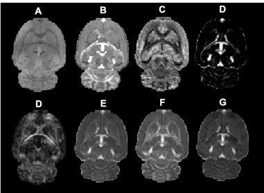
Figure 2 - Single-subject axial slices of A) raw diffusion data B) Neurite Density Index (NDI), C) Orientation Dispersion Index (ODI), D) Isotropic Volume Fraction (IsoVF), E) Fractional Anisotropy (FA), F) Mean Diffusivity (MD), G) Axial Diffusivity H), Radial Diffusivity.
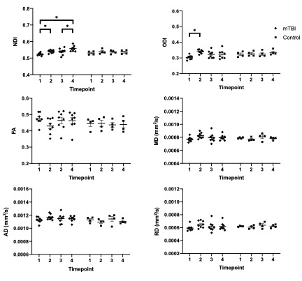
Figure 3 – Mean ± SEM metrics within the corpus callosum for injured and control subjects. Note, injury 1 took place at timepoint 1 and injury 2 took place at timepoint 4. For NDI and ODI, a repeated measures ANOVA was used to determine individual differences amongst the injured and control groups. Post-hoc pairwise comparisons (Bonferroni corrected) were then determined between individual timepoints within each group. Statistically significant (p < .05) pairwise differences are indicated by *.