Yujian Diao1,2,3, Rolf Gruetter3, and Ileana O. Jelescu1,2
1CIBM Center for Biomedical Imaging, Lausanne, Switzerland, 2Animal Imaging and Technology, Ecole Polytechnique Fédérale de Lausanne, Lausanne, Switzerland, 3Laboratory of Functional and Metabolic Imaging, Ecole Polytechnique Fédérale de Lausanne, Lausanne, Switzerland
1CIBM Center for Biomedical Imaging, Lausanne, Switzerland, 2Animal Imaging and Technology, Ecole Polytechnique Fédérale de Lausanne, Lausanne, Switzerland, 3Laboratory of Functional and Metabolic Imaging, Ecole Polytechnique Fédérale de Lausanne, Lausanne, Switzerland
Dynamic functional connectivity in a rat model of Alzheimer’s was marked
by early hyper-connectivity vs control rats, and a decline of specific brain
states over time – both consistent with static FC analysis.
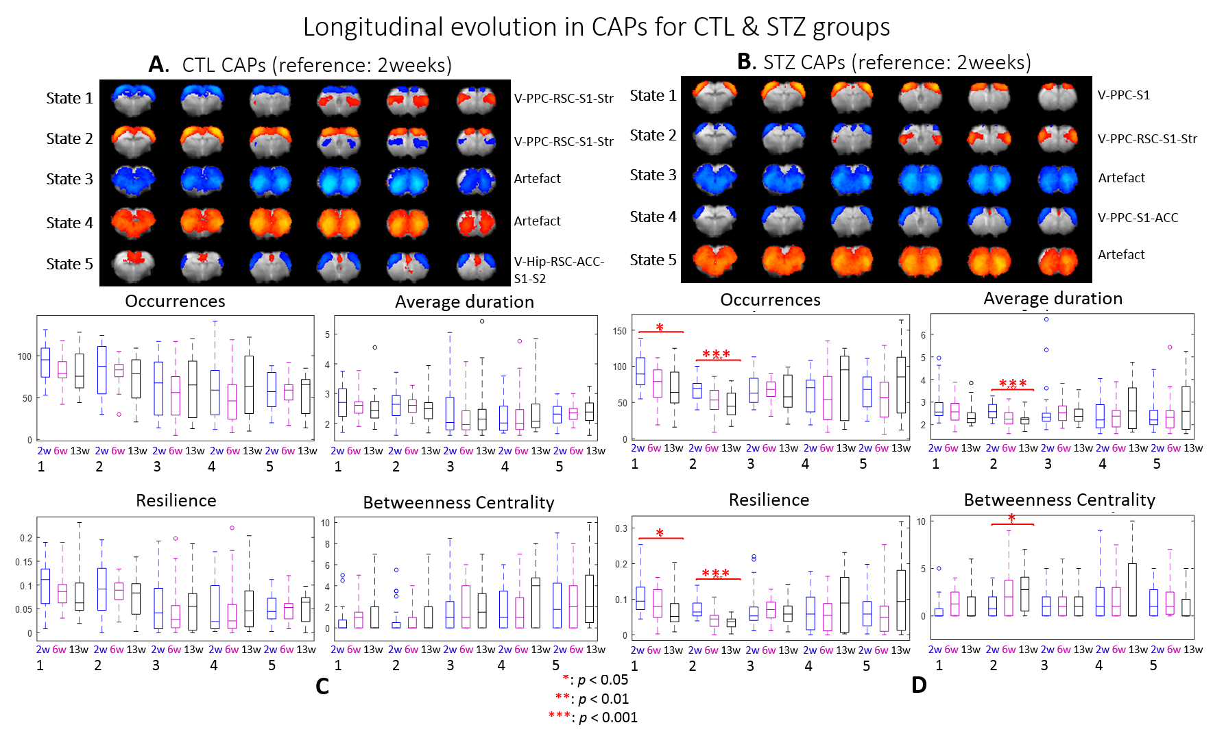
Fig. 5. Longitudinal evolution of CAPs for each group. A & B: 5 main CAPs based on the 2-week data for CTL & STZ group,
respectively. C & D: longitudinal changes in 4 metrics of
the 5 CAPs for CTL and STZ group, respectively. In CTL rats, no significant
difference was detected with time. However, major longitudinal differences were
found in the STZ group. CAP 1 & 2 occurred increasingly less and tended to become
transit states with short duration, resilience and higher betweenness. Those CAPs
cover mostly visual and somatosensory cortex, PPC, RSC and striatum.
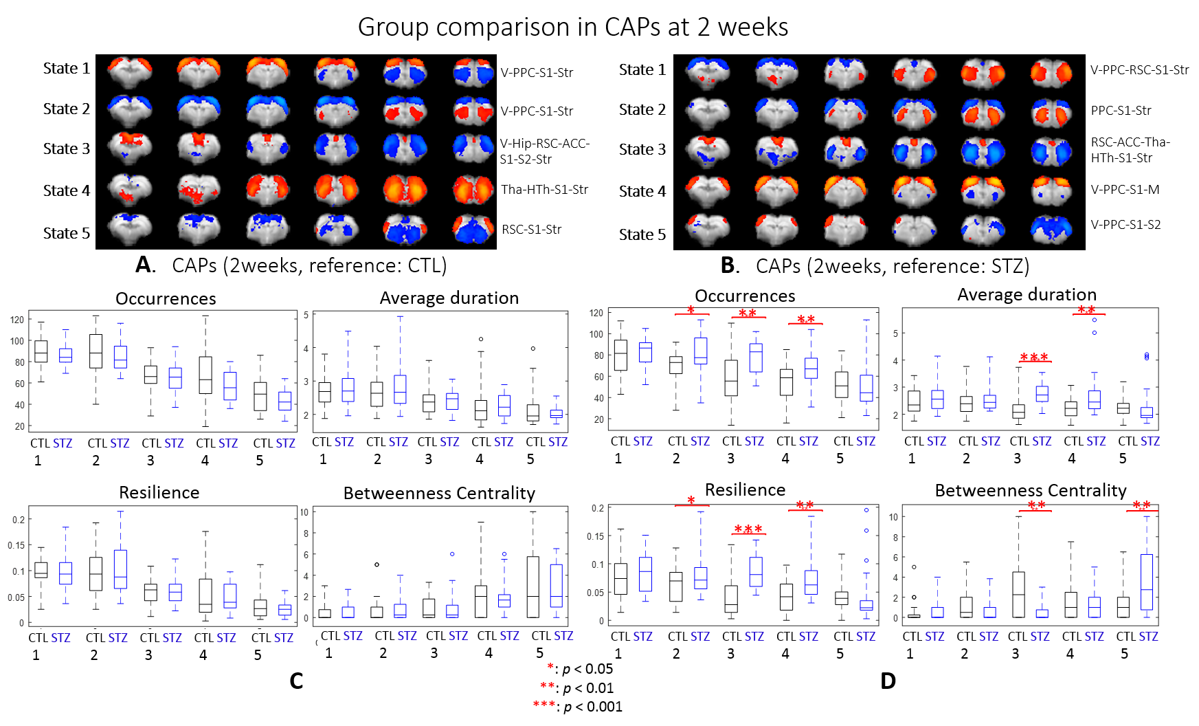
Fig. 2. Intergroup comparison of CAPs at 2 weeks. A & B: 5 main CAPs based
on CTL & STZ, respectively. C &
D: intergroup differences in 4
metrics for CAPs in A & B, respectively. No significant differences were
found when CTL was the reference (C) while the main CAPs 2, 3 & 4 identified
from the STZ group (D) had significantly more occurrences and higher resilience
than their counterpart in CTL. These CAPs mainly cover RSC, PPC, ACC, visual,
motor and somatosensory cortex, thalamus, hypothalamus and striatum.