Andreea Hertanu1,2, Cem Karakus3,4, Lucas Soustelle1,2, Victor N. D. Carvalho1,2,5, Gopal Varma6, David C. Alsop6, Bilal El Waly3,4, Olivier M. Girard1,2, Franck Debarbieux3,4, and Guillaume Duhamel1,2
1Aix Marseille Univ, CNRS, CRMBM, Marseille, France, 2APHM, Hôpital Universitaire Timone, CEMEREM, Marseille, France, 3Aix Marseille Univ, CNRS, INT, Marseille, France, 4Aix Marseille Univ, CNRS, CERIMED, Marseille, France, 5Aix Marseille Univ, CNRS, ICR, Marseille, France, 6Division of MR Research, Radiology, Beth Israel Deaconess Medical Center, Harvard Medical School, Boston, MA, United States
1Aix Marseille Univ, CNRS, CRMBM, Marseille, France, 2APHM, Hôpital Universitaire Timone, CEMEREM, Marseille, France, 3Aix Marseille Univ, CNRS, INT, Marseille, France, 4Aix Marseille Univ, CNRS, CERIMED, Marseille, France, 5Aix Marseille Univ, CNRS, ICR, Marseille, France, 6Division of MR Research, Radiology, Beth Israel Deaconess Medical Center, Harvard Medical School, Boston, MA, United States
The
rostro-caudal ihMTR profile in the LPC-incubated spinal cord follows the
dynamics of demyelination and axonal loss measured by microscopy. T1Ds,
T1 and T2 quantification in the lesion showed different values
than those obtained in the ventral WM taken as reference.
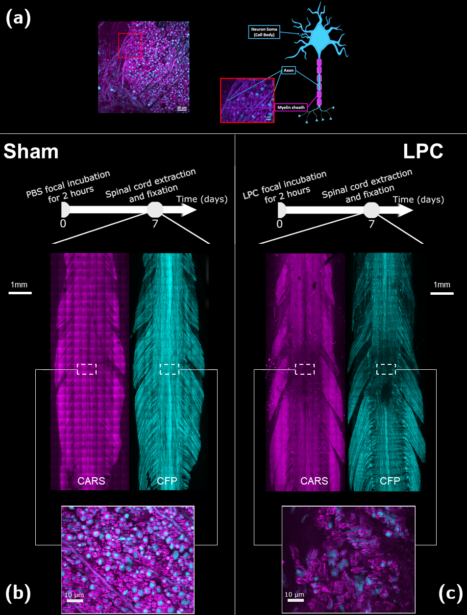
Figure 1:
Schematic depiction of two CARS (magenta) and axonal (cyan) signals (a) and
representative axial and coronal Maximum Intensity Projection views of the
healthy (b) and demyelinated (c) spinal cords with CARS (myelin, magenta) and
Thy1–CFP fluorescence (axons, cyan) contrasts.
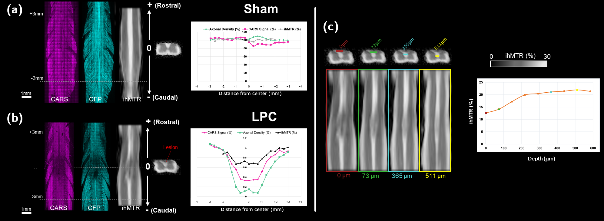
Figure
2: Rostro-caudal profiles of ihMTR, CARS signal and axonal density normalized
to the mean of their values at +3 mm and -3 mm from the lesion, measured in (a)
PBS-incubated spinal cord and (b) LPC-incubated spinal cord. (c) Dorso-ventral
profile of absolute ihMTR in the triangle of WM dorsal tracts.
