Haimei Cao1, Xiang Xiao1, Jun Hua2,3, Guanglong Huang4, Xiaodan Li1, Wenle He1, Jie Qin1, and Yuankui Wu1
1Department of Medical Imaging, Nanfang Hospital, Southern Medical University, Guangzhou, China, 2Neurosection, Division of MRI Research, Department of Radiology, Johns Hopkins University School of Medicine, Baltimore, MD, United States, 3F.M. Kirby Research Center for Functional Brain Imaging, Kennedy Krieger Institute, Department of Radiology, Johns Hopkins University School of Medicine, Baltimore, MD, United States, 4Department of Neurosurgery, Nanfang Hospital, Southern Medical University, Guangzhou, China
1Department of Medical Imaging, Nanfang Hospital, Southern Medical University, Guangzhou, China, 2Neurosection, Division of MRI Research, Department of Radiology, Johns Hopkins University School of Medicine, Baltimore, MD, United States, 3F.M. Kirby Research Center for Functional Brain Imaging, Kennedy Krieger Institute, Department of Radiology, Johns Hopkins University School of Medicine, Baltimore, MD, United States, 4Department of Neurosurgery, Nanfang Hospital, Southern Medical University, Guangzhou, China
Both iVASO-rCBVa
and DWI-mADC can predict gliomas grades and combining these two parameters can
further improve diagnostic performance. Also, VASO and DWI have the added value
to structual MRI in preoperative prediction of tumor grading of gliomas.
Figure
2. Right frontal lobe anaplastic oligodendroglioma in a 51-year-old male (Grade
III). The lesion in the right frontal lobe was iso-intense with perilesional
edema on T2WI (A) and showed no enhancement on contrast-enhanced T1WI (B). The
lesion showed an iso-intensity on diffusion-weighted image (C) and the focus of
elevated arteriolar perfusion (arrowhead) on CBVa map (D). On review 1, the
lesion was diagnosed as a low-grade glioma. On review 2, the lesion was
diagnosed as a high-grade glioma.
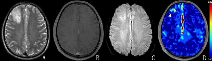
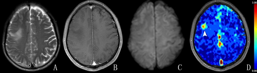
Figure 1. Right frontal lobe anaplastic astrocytoma
(Grade III) in a 39-year-old male. The lesion in the right frontal lobe was
hyper-intense on T2WI (A) and showed no enhancement on contrast-enhanced T1WI
(B). The lesion showed a slight hyper-intensity on diffusion-weighted image (C)
and the focus of elevated arteriolar perfusion (arrowhead) on CBVa map (D). On
review 1, the lesion was diagnosed as a low-grade glioma. On review 2, the
lesion was diagnosed as a high-grade glioma.