Laleh Eskandarian1,2, Safak Parlak3, Onur Afacan4,5, Ceren Günbey6, Nesibe Gevher Ertuğrul6, Banu Anlar6, and Kader Karli Oguz2,3
1Neuroscience Department, Bilkent University, Ankara, Turkey, 2National Magnetic Resonance Research Center (UMRAM), Bilkent University, Ankara, Turkey, 3Faculty of Medicine, Department of Radiology, Hacettepe University, Ankara, Turkey, 4Department of Radiology, Boston Children’s Hospital, Boston, MA, United States, 5Department of Radiology, Harvard Medical School, Boston, MA, United States, 6Department of Pediatrics, Hacettepe University, Ankara, Turkey
1Neuroscience Department, Bilkent University, Ankara, Turkey, 2National Magnetic Resonance Research Center (UMRAM), Bilkent University, Ankara, Turkey, 3Faculty of Medicine, Department of Radiology, Hacettepe University, Ankara, Turkey, 4Department of Radiology, Boston Children’s Hospital, Boston, MA, United States, 5Department of Radiology, Harvard Medical School, Boston, MA, United States, 6Department of Pediatrics, Hacettepe University, Ankara, Turkey
MLD is a dysmyelinating autosomal recessive lysosomal storage disease. T2WI may not show the disease involvement accurately, especially in early phases where a bone marrow transplant can be a treatment option. We used Myelin Water Fraction and DTI to investigate WM in patients with MLD.
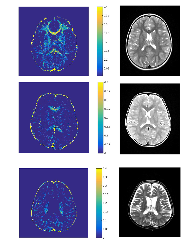
Figure 1. Axial T2WI and
T2 MWF maps
of a
7-year old female HC (A)
and
two
patients with MLD, 4 and 9-years old at
(B, C respectively)
with
varying
severity
of leukodystrophy.
Both
patients
have
reduced
MWF compared
with
HC in A. Although
slight
hyperintensity
is noticed
on T2WI; the
patients
have
markedly
low
myelin
on MWF.
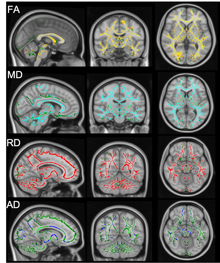
Figure 2. Shows TBSS maps
derived
from
DTI studies
from
the
patients
and
HCs.
Compared
with
HCs,
FA map
shows
widespread
significant
reduction
(A), MD and
RD maps
show
increase
(B and
C ) and
AD map
shows
a limited
reduction
in WM of the
patients.