Shin Tai Chong1, Xinrui Liu2, Hung-Wen Kao3, Chien-Yuan Eddy Lin4, Sanford PC Hsu5, and Ching-Po Lin1
1Institute of Neuroscience, National Yang-Ming University, Taipei, Taiwan, 2Department of Neurosurgery, First Hospital of Jilin University, Jilin, China, 3Tri-Service General Hospital, National Defense Medical Center, Taipei, Taiwan, 4GE Healthcare, Taipei, Taiwan, 5Department of Neurosurgery, Neurological Institute, Taipei Veterans General Hospital, Taipei, Taiwan
1Institute of Neuroscience, National Yang-Ming University, Taipei, Taiwan, 2Department of Neurosurgery, First Hospital of Jilin University, Jilin, China, 3Tri-Service General Hospital, National Defense Medical Center, Taipei, Taiwan, 4GE Healthcare, Taipei, Taiwan, 5Department of Neurosurgery, Neurological Institute, Taipei Veterans General Hospital, Taipei, Taiwan
Neurite Orientation Dispersion and Density Imaging (NODDI)-based tractography could improve the reconstruction of the fiber tracts that by tracking through regions of peritumoral edema.
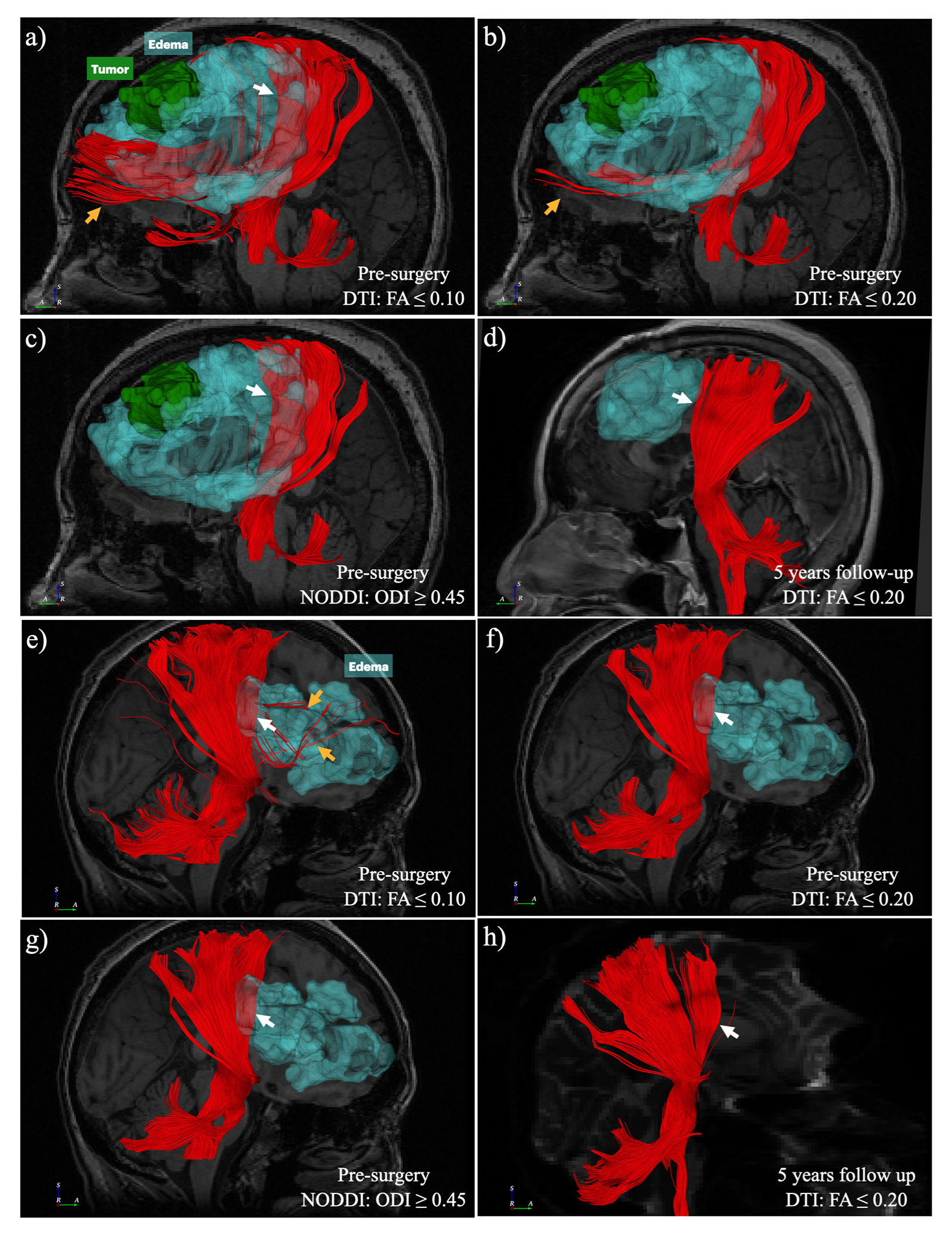
Two examples (a-d, meningioma, WHO grade II; e-h, malignant B-cell lymphoma) of visual comparison between DTI and NODDI-based CST reconstruction. NODDI-based tractography with ODI has better performance in the region of peritumoral edema and fewer false-positive tracts (c & g, white arrow). After the vasogenic edema subsided, the DTI-based tractography with FA ≤ 0.2 shows CST similar to the pre-surgery NODDI-based CST (d & h).

NODDI's volume fraction distributions in the DN group and N group are plotted with a 2D barycentric coordinate system. As compared with the DN group (a), the N group (b) distribution shows a left upper shift in the coordinate system. There is no significant difference in VFec (P = 0.733, d), but significant differences in VFic (P < 0.001, e) and VFiso (P = 0.025, c).