Weike Zeng1 and Mengzhu Wang2
1Sun Yat-sen Memorial Hospital, Sun Yat-sen University, Guangzhou, China, 2MR Scientific Marketing, Siemens Healthcare, Guangzhou, China
1Sun Yat-sen Memorial Hospital, Sun Yat-sen University, Guangzhou, China, 2MR Scientific Marketing, Siemens Healthcare, Guangzhou, China
Decreased ICVF value in brain edema lesion indicated the reduction of neurite density, and was associated with cognitive decline in RI. NODDI as a new MRI diffusion technique may contribute to better understanding of pathophysiology of cognitive decline in RI than DTI.
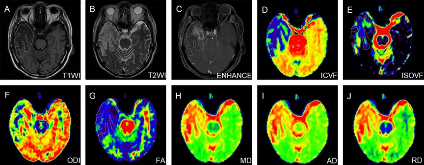
Figure 1. NODDI maps of a patient with RI, together with DTI maps, T1WI, T2WI and enhanced images. RI, radiation-induced brain injury; T1WI, T1-weighted imaging; T2WI, T2-weighted imaging; FA, fractional anisotropy; MD, mean diffusivity; AD, axial diffusivity; RD, radial diffusivity; ICVF, intra-neurite volume fraction; ODI, orientation dispersion index; ISOVF, volume fraction of the isotropic compartment.
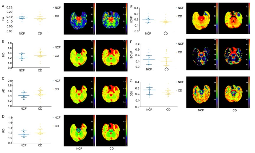
Figure 2. Differences of DTI parameters including FA (A), MD (B), RD (C) and MD (D) as well as NODDI parameters including ICVF (E), ISOVF (F) and ODI (G) of edema lesions between patients with or without cognitive decline in RI. RI, radiation- induced brain injury; NCF, normal cognitive function; CD, cognitive decline; FA, fractional anisotropy; MD, mean diffusivity. AD, axial diffusivity; RD, radial diffusivity; ICVF, intra-neurite volume fraction; ODI, orientation dispersion index; ISOVF, volume fraction of the isotropic compartment.