Augusto Lio M. Goncalves Filho1,2, Azadeh Tabari1,2, Chanon Ngamsombat3, Ilena George4, Stephen F. Cauley2, Wei Liu5, Daniel N. Splitthoff6, Wei-Ching Lo7, Pamela W. Schaefer1, Otto Rapalino1, Eric C. Klawiter4, John Conklin1,2, and Susie Y. Huang1,2
1Department of Radiology, Massachusetts General Hospital, Boston, MA, United States, 2Department of Radiology, Athinoula A. Martinos Center for Biomedical Imaging, Charlestown, MA, United States, 3Department of Radiology, Siriraj Hospital, Bangkok, Thailand, 4Department of Neurology, Massachusetts General Hospital, Boston, MA, United States, 5Siemens Shenzhen Magnetic Resonance Ltd., Shenzhen, China, 6Siemens Healthcare GmbH, Erlangen, Germany, 7Siemens Medical Solutions Inc., Boston, MA, United States
1Department of Radiology, Massachusetts General Hospital, Boston, MA, United States, 2Department of Radiology, Athinoula A. Martinos Center for Biomedical Imaging, Charlestown, MA, United States, 3Department of Radiology, Siriraj Hospital, Bangkok, Thailand, 4Department of Neurology, Massachusetts General Hospital, Boston, MA, United States, 5Siemens Shenzhen Magnetic Resonance Ltd., Shenzhen, China, 6Siemens Healthcare GmbH, Erlangen, Germany, 7Siemens Medical Solutions Inc., Boston, MA, United States
Accelerated Wave-CAIPI susceptibility weighted imaging (SWI) and
FLAIR may improve the characterization of demyelinating lesions within
reasonable acquisition times and provide a more confident diagnosis of multiple
sclerosis in brain MRI at 3T.
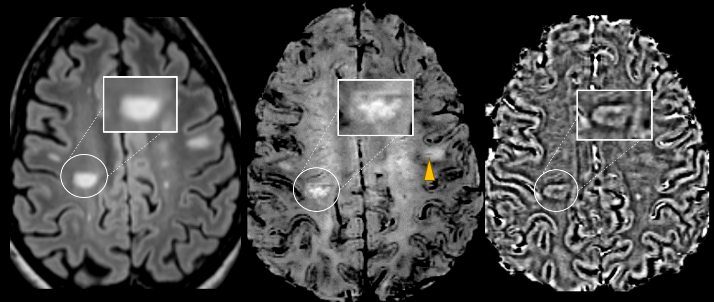
Figure 1. Axial Wave-SPACE-FLAIR
(left), Wave-SWI (middle), and phase images (right) in a 28-year-old
patient with confirmed MS demonstrating the presence of paramagnetic rims
corresponding to lesions visible on FLAIR (highlighted). A lesion with a central
vein sign can be seen in the same Wave-SWI sequence (arrowhead).
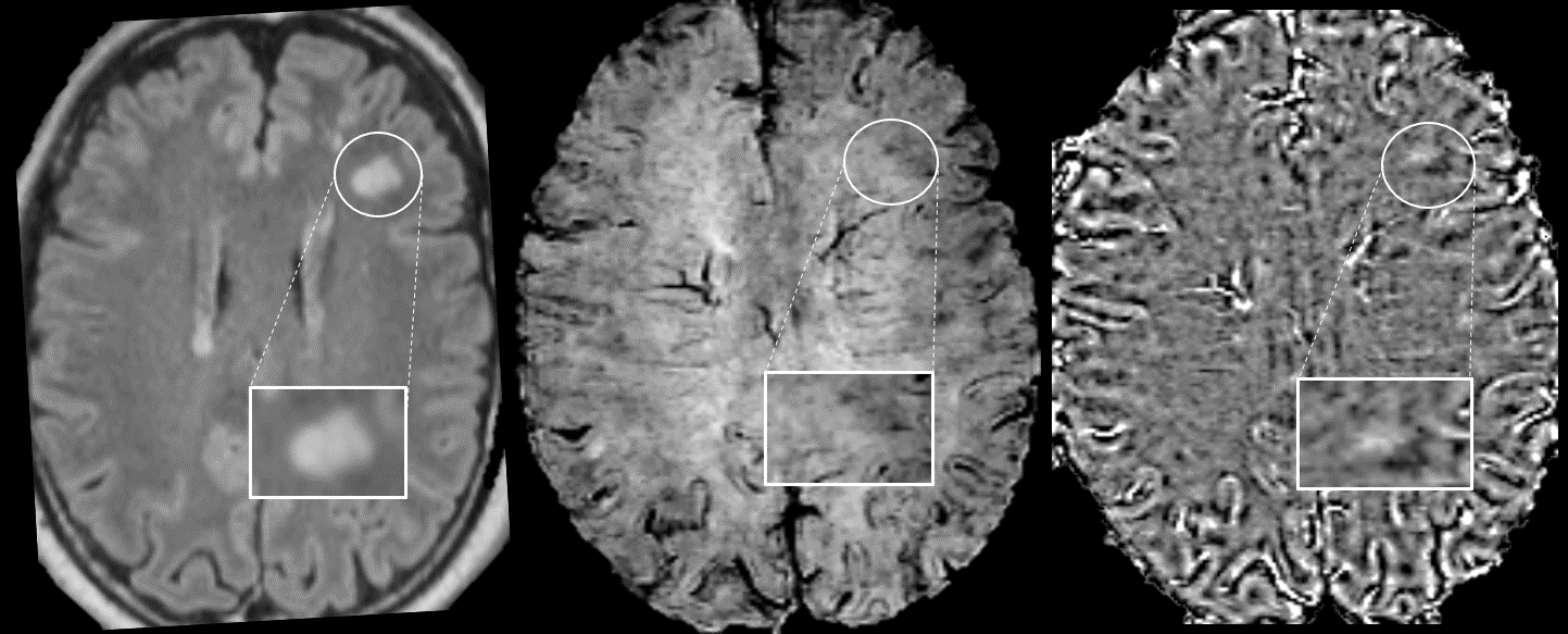
Figure 3. Axial Wave-SPACE-FLAIR
(left), Wave-SWI (middle), and phase images (right) in a 20-year-old
patient with confirmed anti-MOG disease with no visible paramagnetic rim around
the lesion visible on FLAIR (highlighted). No lesions with a central vein sign were
identified on the Wave-FLAIR and Wave-SWI sequences.