Yuankui Wu1, Wenle He1,2, Wensheng Wang2, Jun Hua3,4, Xiang Xiao1, Xiaomin Liu1, Yikai Xu1, and Yingjie Mei5
1Department of Medical Imaging, Nanfang Hospital, Southern Medical University, Guangzhou, China, 2Department of Radiology, Guangdong 999 Brain Hospital, Guangzhou, China, 3Neurosection, Division of MRI Research, Department of Radiology, Johns Hopkins University School of Medicine, Baltimore, MD, United States, 4F.M. Kirby Research Center for Functional Brain Imaging, Kennedy Krieger Institute, Department of Radiology, Johns Hopkins University School of Medicine, Baltimore, MD, United States, 5Philips healthcare, Guangzhou, China
1Department of Medical Imaging, Nanfang Hospital, Southern Medical University, Guangzhou, China, 2Department of Radiology, Guangdong 999 Brain Hospital, Guangzhou, China, 3Neurosection, Division of MRI Research, Department of Radiology, Johns Hopkins University School of Medicine, Baltimore, MD, United States, 4F.M. Kirby Research Center for Functional Brain Imaging, Kennedy Krieger Institute, Department of Radiology, Johns Hopkins University School of Medicine, Baltimore, MD, United States, 5Philips healthcare, Guangzhou, China
The top two CBVa histogram features in predicting MGMT promoter methylation
in grade II – IV gliomas were RMS & Varianc. Combing CBVa histogram
features and structural MRI features improved diagnostic performance.
Figure
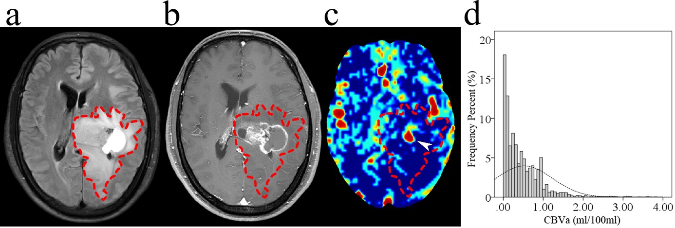
1. Glioblastoma with MGMT promoter methylation in a 39-year-old male. (a) Axial
T2-FLAIR image. (b) Axial postcontrast T1-weighted image. (c) Axial CBVa map
(ml/100 ml). (d) Histogram of CBVa from whole tumor ROI (dashed line). Images
show a left thalamic mass with cystic change, which involved the lateral
ventricular walls but spared the cortices. The CBVa map shows focal
hyperperfusion (arrowhead) in the mass. The corresponding histogram shows a narrow
distribution of perfusion in the whole tumor which is mainly concentrated in hypoperfused
areas.
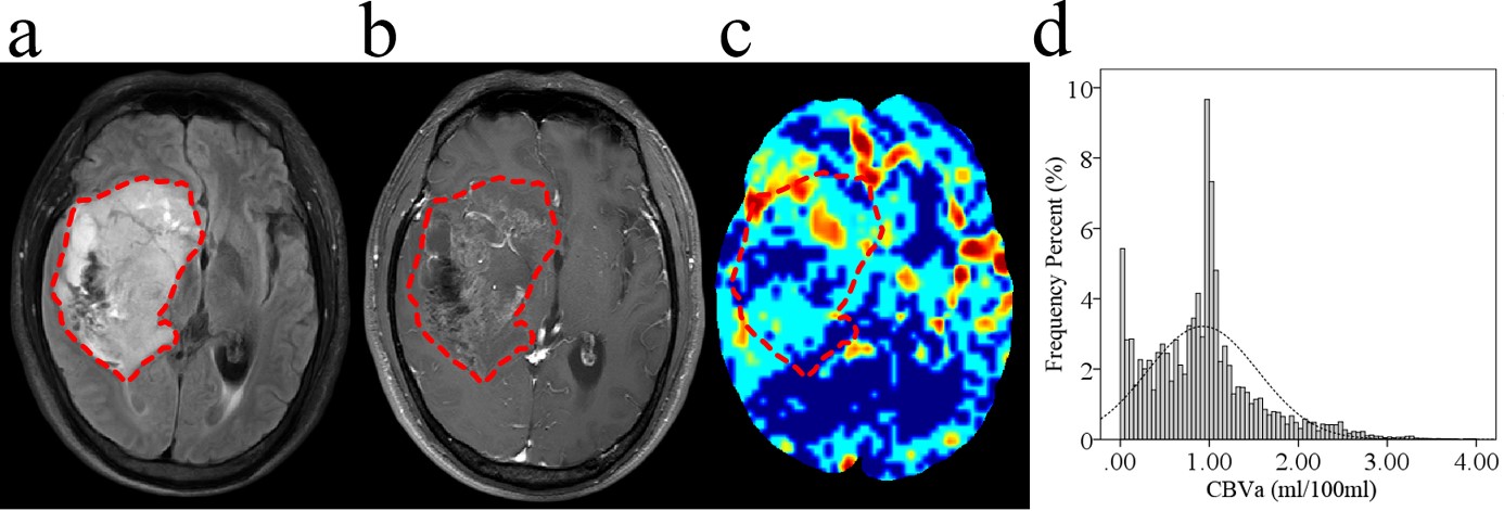
Figure 2. Glioblastoma
without MGMT promoter methylation in a 44-year-old male. (a) Axial T2-FLAIR
image. (b) Axial postcontrast T1-weighted image. (c) Axial CBVa map (ml/100
ml). (d) Histogram of CBVa from whole tumor ROI (dashed line). The images
show an inhomogeneous mass in the right temporal lobe, which invaded the right
basal ganglia, the cortices, and the subventricular zone. The corresponding
histogram shows a wide distribution of perfusion in the whole tumor, with a
peak located at around 1.0 ml/100 ml.