Jason Langley1, Sana Hussain2, Daniel E Huddleston3, Ilana Bennett4, and Xiaoping P Hu1,2
1Center for Advanced Neuroimaging, University of California Riverside, Riverside, CA, United States, 2Department of Bioengineering, University of California Riverside, Riverside, CA, United States, 3Department of Neurology, Emory University, Atlanta, GA, United States, 4Department of Psychology, University of California Riverside, Riverside, CA, United States
1Center for Advanced Neuroimaging, University of California Riverside, Riverside, CA, United States, 2Department of Bioengineering, University of California Riverside, Riverside, CA, United States, 3Department of Neurology, Emory University, Atlanta, GA, United States, 4Department of Psychology, University of California Riverside, Riverside, CA, United States
Locus coeruleus axons
project to the thalamus via the central tegmental tract (CTT). In the APOE-e4 positive group, CTT
microstructural measures were positively correlated with thalamus
tau-PET SUVR but no correlations were observed in the APOE-e4 negative group.
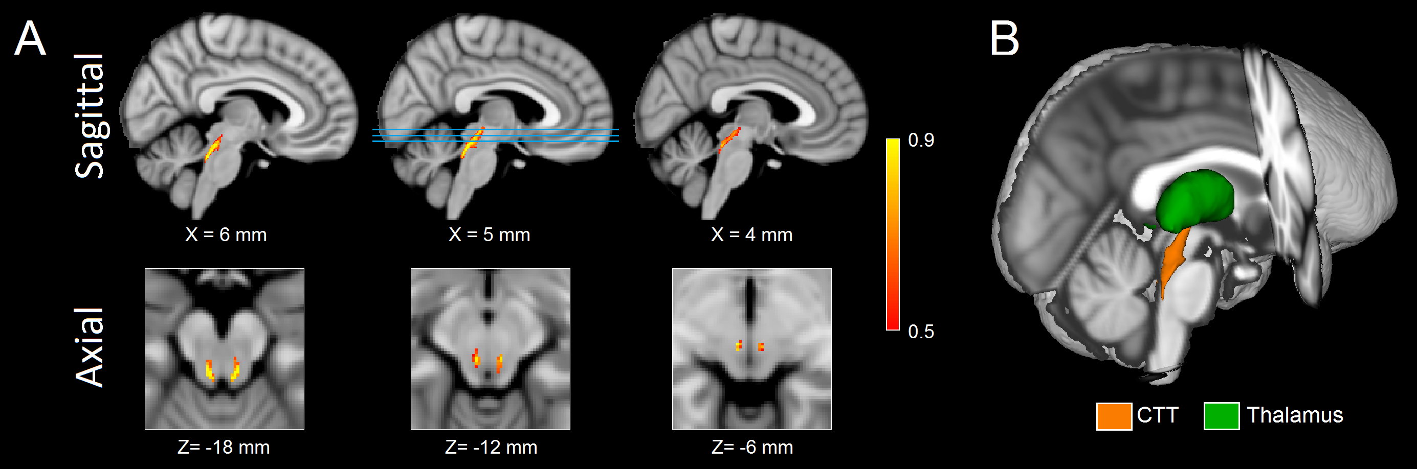
Figure 1. Sagittal (A; top row) and axial (A; bottom row) of population maps showing the CTT ROI used in this analysis. The lines in sagittal view at X=5 mm identify the location of the axial slices shown in the bottom row in MNI space. A three dimensional rendering of the CTT is shown in B.
CTT - central tegmental tract; ROI - region of interest; MNI - Montreal Neurological Institute
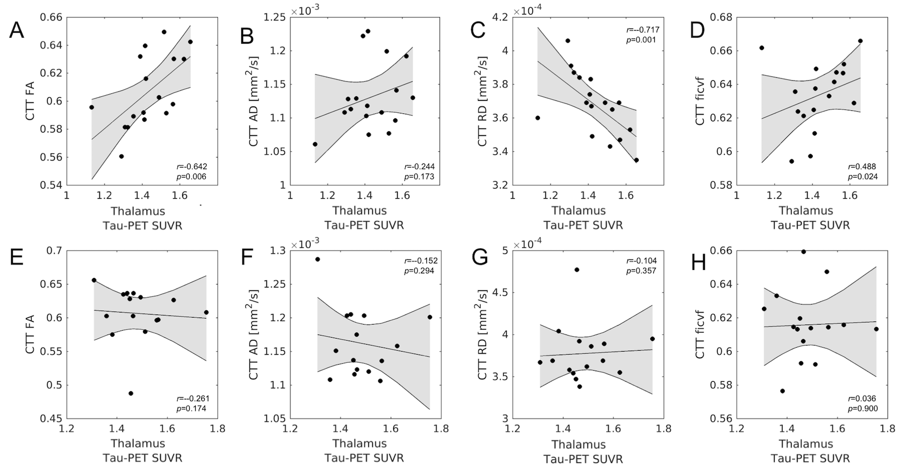
Figure 3. Correlations between CTT microstructure metrics and thalamus
tau-PET SUVR in APOE-ε4 positive subjects (top row; A-D) and APOE-ε4 negative subjects (bottom row; E-H). Significant
correlations between CTT microstructure and thalamus tau-PET SUVR are seen in
the APOE-ε4 positive group but not in the APOE-ε4 negative group.