Alexa Haeger1,2,3, Michel Bottlaender1,4, Julien Lagarde4,5,6, Renata Porciuncula Baptista1, Cécile Rabrait-Lerman1, Volker Luecken2,3, Jörg Bernhard Schulz2,3, Alexandre Vignaud1, Marie Sarazin4,5,6, Kathrin Reetz2,3, Sandro Romanzetti2,3, and Fawzi Boumezbeur1
1BAOBAB, CNRS, Paris-Saclay University, CEA-NeuroSpin, Gif-sur-Yvette, France, 2Department of Neurology, RWTH Aachen University, Aachen, Germany, 3JARA-BRAIN Institute of Molecular Neuroscience and Neuroimaging, Forschungszentrum Jülich GmbH, Julich, Germany, 4BioMaps, CNRS, Inserm, Paris-Saclay University, CEA-SHFJ, Orsay, France, 5Neurology of Memory and Language, GHU Paris Psychiatrie & Neurosciences, Sainte-Anne Hospital, Paris, France, 6Université de Paris, Paris, France
1BAOBAB, CNRS, Paris-Saclay University, CEA-NeuroSpin, Gif-sur-Yvette, France, 2Department of Neurology, RWTH Aachen University, Aachen, Germany, 3JARA-BRAIN Institute of Molecular Neuroscience and Neuroimaging, Forschungszentrum Jülich GmbH, Julich, Germany, 4BioMaps, CNRS, Inserm, Paris-Saclay University, CEA-SHFJ, Orsay, France, 5Neurology of Memory and Language, GHU Paris Psychiatrie & Neurosciences, Sainte-Anne Hospital, Paris, France, 6Université de Paris, Paris, France
By combining quantitative 23Na MRI at 7
Tesla with Tau/Amyloid-PET, we show that total sodium concentration is
increased in AD patients compared to controls and that these changes are more strongly
correlated with local Tau- than Amyloid-loads.
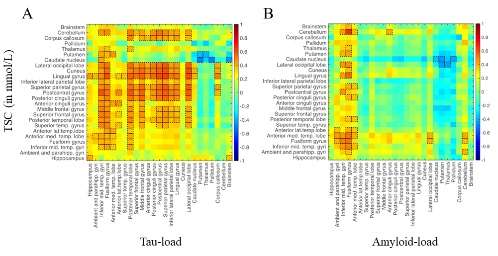
Figure 4. Correlation matrices between TSC versus local Tau
load (A) and versus local Amyloid load (B). Notable correlations
are reported at an uncorrected p < 0.05 level of significance and are framed
in black.
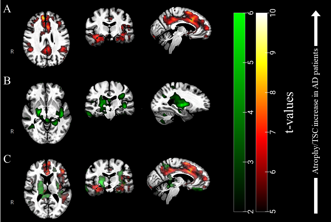
Figure 3. (A) T-values resulting
from voxel-based permutation analyses of our TSC maps (AD > Controls) and (B) atrophy clusters in green (Controls
> AD), overlaid to our group template. (C) From the overlay of both TSC and
atrophy clusters, one can appreciate the differences between both pathological patterns.