Fan Yang1, Jian Zhang2, Guobin Li2, Jiayu Zhu2, Xin Tang1, and Chenxi Hu1
1Institute of Medical Imaging Technology, School of Biomedical Engineering, Shanghai Jiao Tong University, Shanghai, China, 2United Imaging Healthcare Co., Ltd, Shanghai, China
1Institute of Medical Imaging Technology, School of Biomedical Engineering, Shanghai Jiao Tong University, Shanghai, China, 2United Imaging Healthcare Co., Ltd, Shanghai, China
Prospective SUPER-CAIPIRINHA
shows consistent image quality and accurate T1 quantification compared with
retrospective SUPER-CAPIRINHA and the gold standard. The scan time over the
entire cerebrum is reduced from more than 6 minutes to 1.5 mintues.
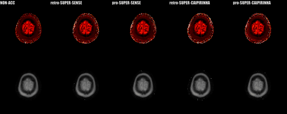
Figure
2. Reconstructed 3D T1 and M0 maps of whole cerebrum
for 1 healthy subject. Image quality was
similar
between SUPER-SENSE and SUPER-CAIPIRINHA and between retrospective and
prospective reconstruction. Image
fine details were truthfully
preserved even for 5-fold acceleration. SUPER-SENSE
and SUPER-CAIPIRINHA reduced the scan time from 6:11 minutes to 2:09 minutes
and 1:29 minutes, respectively.
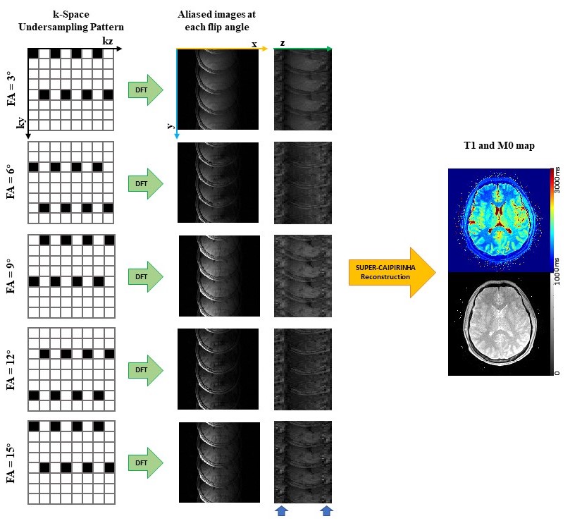
Figure
1. Acceleration pipeline of SUPER-CAIPIRINHA for 5-fold acceleration.
Column
1 shows k-Space undersampling
pattern of SUPER-CAIPIRINHA, black blocks represent points to be sampled while
white blocks represent undersampled
points in k-space.
Severe aliasing was caused by high undersampling
rate (column 2&3). Parametric maps
without aliasing artifact
were reconstructed by blockwise
curve-fitting method of SUPER (column
4).