Nicholas Dwork1, Daniel O'Connor2, Corey A. Baron3, Ethan M. I. Johnson4, John M. Pauly5, and Peder E.Z. Larson6
1Radiology and Biomedical Imaging, UCSF, San Francisco, CA, United States, 2Mathematics and Statistics, University of San Francisco, San Francisco, CA, United States, 3Robarts Research, Western University, London, ON, Canada, 4Biomedical Engineering, Northwestern University, Evanston, IL, United States, 5Electrical Engineering, Stanford University, Stanford, CA, United States, 6Radiology and Biomedical Imaging, University of California in San Francisco, San Francisco, CA, United States
1Radiology and Biomedical Imaging, UCSF, San Francisco, CA, United States, 2Mathematics and Statistics, University of San Francisco, San Francisco, CA, United States, 3Robarts Research, Western University, London, ON, Canada, 4Biomedical Engineering, Northwestern University, Evanston, IL, United States, 5Electrical Engineering, Stanford University, Stanford, CA, United States, 6Radiology and Biomedical Imaging, University of California in San Francisco, San Francisco, CA, United States
The structure of the wavelet transform is used to improve results from compressed sensing.
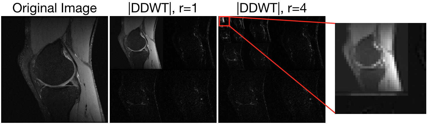
(Left) original MR image of a knee. (Center-left) Daubechies Wavelet transform of the knee. (Center-right) Daubechies Wavelet transform of the knee with $4$ levels of recursion. (Right) Zoom in of lowest-frequency portion shows a lack of sparsity.
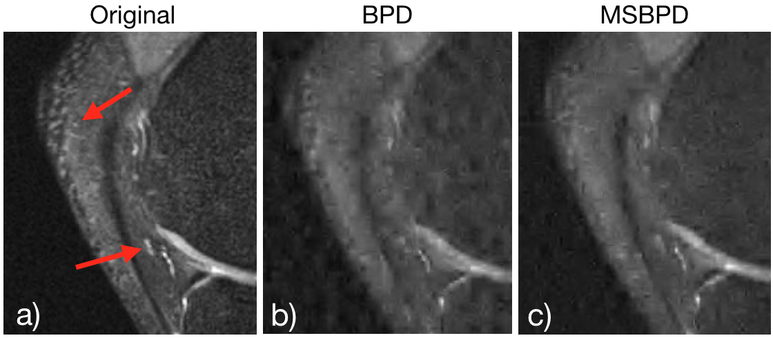
Zoom in to reconstructions of MRI data of knee from 8% of the data. (a) shows the original image; (b) shows the reconstruction using BPD; and (c) shows reconstruction with MSBPD. The red arrows point to regions in the imagery where the improvement in quality of MSBPD over the other algorithms is very apparent. Furthermore, one notes that the variations in the bone marrow are significantly less in the MSBPD reconstruction than in the BPD result, as expected.