Corey Edward Cruttenden1, Wei Zhu1, Yi Zhang1, Xiao-Hong Zhu1, Rajesh Rajamani2, and Wei Chen1
1Center for Magnetic Resonance Research, University of Minnesota, Minneapolis, MN, United States, 2Mechanical Engineering, University of Minnesota, Minneapolis, MN, United States
1Center for Magnetic Resonance Research, University of Minnesota, Minneapolis, MN, United States, 2Mechanical Engineering, University of Minnesota, Minneapolis, MN, United States
The first difference of fMRI-artifact-contaminated neuro-electrophysiology is separable into the clean neural signals and artifact contributions by singular value shrinkage. The method is demonstrated on neural signals acquired during EPI at 16.4T.

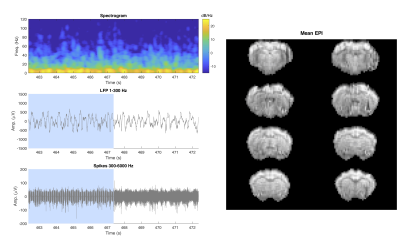
Spectrogram (top left) and local field potential (middle left) during and immediately after EPI scan. The EPI scan period (up to 467.6 s) is indicated by blue shading in the time-domain plots. Neuronal spike signal during the same period (bottom left). The SVD shrinkage approach uncovers excellent quality LFP recordings when compared to post-scan time periods in the spectrogram and time domain. The mean EPI images acquired during the recording are presented on the right.

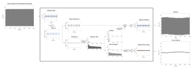
Schematic of artifact removal approach. Artifact contaminated data is aligned by TR and the first difference is computed, followed by mean waveform separation, and SVD-based shrinkage on residuals to further separate artifacts from neural signals. Separated artifact and neural signal contributions are reconstructed and integrated (cumulative summation) to form artifact and clean neural signal estimates. Data acquired from rat cortex in vivo during echo planar imaging at 16.4T.