Rolf F Schulte1, Mary A McLean2, Joshua D Kaggie2, Stephan Ursprung2, Ramona Woitek2, Ferdia A Gallagher2, Esben S S Hansen3, Nikolaj Bogh3, and Christoffer Laustsen3
1GE Healthcare, Munich, Germany, 2Department of Radiology, University of Cambridge, Cambridge, United Kingdom, 3MR Research Centre, University of Aarhus, Aarhus, Denmark
1GE Healthcare, Munich, Germany, 2Department of Radiology, University of Cambridge, Cambridge, United Kingdom, 3MR Research Centre, University of Aarhus, Aarhus, Denmark
SVD-based
coil-combination improves SNR in metabolic imaging of hyperpolarised 13C
compounds and is robust enough to be included in a fully automatic processing
pipeline.
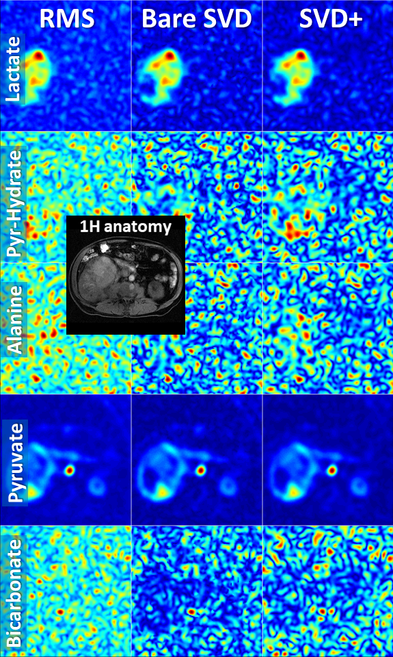
Fig. 1: Comparison of different coil combinations in a kidney cancer patient acquired
with an 8-channel receive coil (average of time steps 3 to 10; slice 3 out of 5
slices).
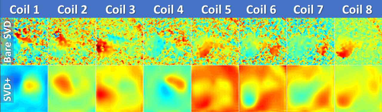
Fig. 2: Sensitivity maps in the same kidney cancer patient as in Fig. 1 (real part only) acquired with eight 13C receive channels: raw receive sensitivities after SVD calculation are shown in the top row (“bare SVD”) and the sensitivities after polynomial smoothing and channel normalisation in the bottom row (“SVD+”).