Ning Jin1, Maria Monzon2, Teodora Chitiboi3, Aaron Pruitt4, Daniel Giese2, Matthew Tong5, and Orlando P Simonetti5,6,7
1Cardiovascular MR R&D, Siemens Medical Solutions USA, Inc., Cleveland, OH, United States, 2Siemens Healthcare, Erlangen, Germany, 3Siemens Medical Solutions USA, Inc, Princeton, NJ, United States, 4Biomedical Engineering, The Ohio State University, Columbus, OH, United States, 5Internal Medicine, The Ohio State University, Columbus, OH, United States, 6Davis Heart & Lung Research Institute, The Ohio State University, Columbus, OH, United States, 7Radiology, The Ohio State University, Columbus, OH, United States
1Cardiovascular MR R&D, Siemens Medical Solutions USA, Inc., Cleveland, OH, United States, 2Siemens Healthcare, Erlangen, Germany, 3Siemens Medical Solutions USA, Inc, Princeton, NJ, United States, 4Biomedical Engineering, The Ohio State University, Columbus, OH, United States, 5Internal Medicine, The Ohio State University, Columbus, OH, United States, 6Davis Heart & Lung Research Institute, The Ohio State University, Columbus, OH, United States, 7Radiology, The Ohio State University, Columbus, OH, United States
We developed a fully automated segmentation algorithm for phase-contrast MR images using deep learning (DL). Automated segmentation of aorta and main pulmonary artery from
PC MRI scans can be successfully achieved using the DL model.
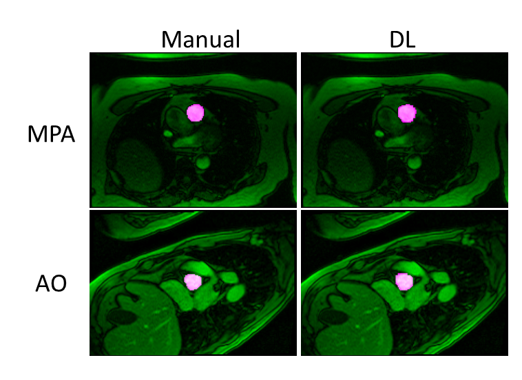
Figure
2. Representative
example of vessel contouring performed by manual and DL segmentation in MPA and
AO.
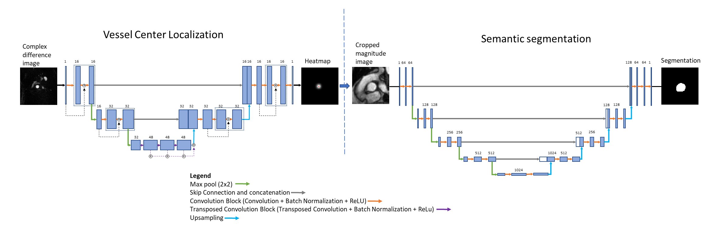
Figure 1.
Schematic representation of the proposed segmentation model. A 2D U-net
model with 3 encoder-decoder blocks is trained to regress heatmaps directly from input complex
difference images to localize vessel center. A second 2D U-net model
with 5 encoder-decoder blocks is trained to segment out the vessel using cropped magnitude
images as input.