Julio A. Oscanoa1,2, Matthew J. Middione2, Christopher M. Sandino3, Shreyas S. Vasanawala2, and Daniel B. Ennis2,4
1Department of Bioengineering, Stanford University, Stanford, CA, United States, 2Department of Radiology, Stanford University, Stanford, CA, United States, 3Department of Electrical Engineering, Stanford University, Stanford, CA, United States, 4Cardiovascular Institute, Stanford University, Stanford, CA, United States
1Department of Bioengineering, Stanford University, Stanford, CA, United States, 2Department of Radiology, Stanford University, Stanford, CA, United States, 3Department of Electrical Engineering, Stanford University, Stanford, CA, United States, 4Cardiovascular Institute, Stanford University, Stanford, CA, United States
2D PC-MRI datasets can be reconstructed with ±5% accuracy for total flow and peak velocity using the proposed Deep Learning based reconstruction methods for acceleration rates up to 8x.
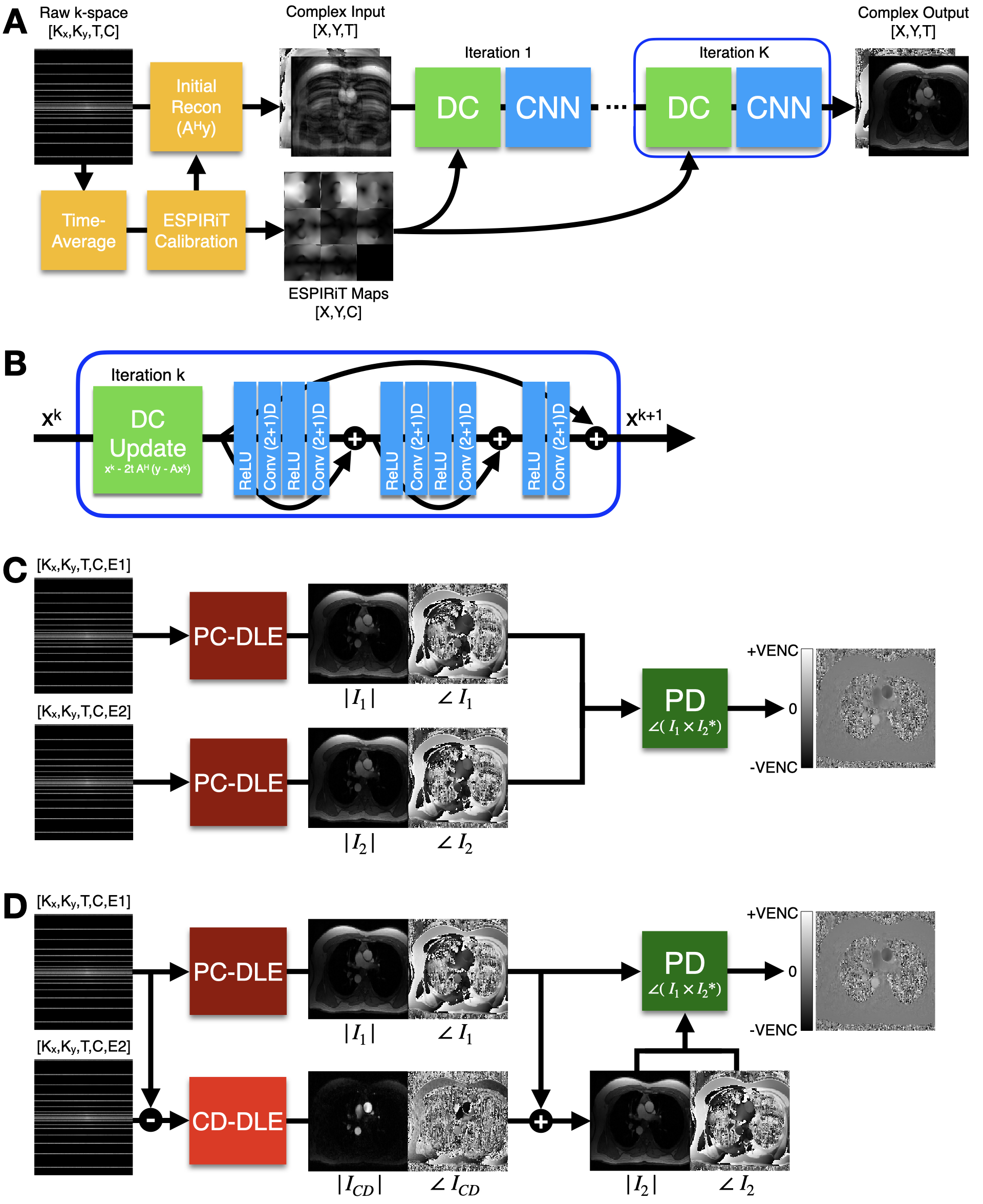
Figure 1. (A) The original DL-ESPIRiT9 reconstruction pipeline with an unrolled network architecture that alternates between a Data Consistency update and a CNN-based denoising step. (B) The CNN was comprised of (2+1)D convolutional layers as described in 9. Both PC-MRI velocity encodings, $$$I_1$$$ and $$$I_2$$$, were used to generate a quantitative phase difference image using either the proposed (C) Phase Contrast DL-ESPIRiT (PC-DLE) or (D) Complex Difference DL-ESPIRiT (CD-DLE) reconstruction pipelines.
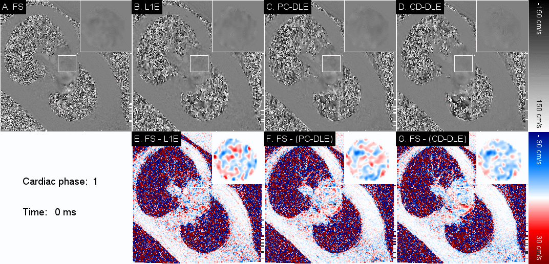
Figure 2. Representative velocity movies for (A) FS and 8x undersampled data reconstructed using (B) L1E, (C) PC-DLE, and (D) CD-DLE. Pixel-by-pixel velocity difference movies are shown for (E) FS vs. L1E, (F) FS vs. PC-DLE, and (G) FS vs. CD-DLE. Both DL frameworks present significantly lower error (F, G), especially at the cardiac phases with high flow.