Haben Berhane1, Michael Scott1, Justin Baraboo1, Cynthia Rigsby2, Joshua Robinson2, Bradley Allen3, Chris Malaisrie3, Patrick McCarthy3, Ryan Avery3, and Michael Markl1
1Biomedical Engineering, Northwestern University, Chicago, IL, United States, 2Lurie Childrens Hospital of Chicago, Chicago, IL, United States, 3Northwestern Radiology, Evanston, IL, United States
1Biomedical Engineering, Northwestern University, Chicago, IL, United States, 2Lurie Childrens Hospital of Chicago, Chicago, IL, United States, 3Northwestern Radiology, Evanston, IL, United States
A
convolutional neural network was trained and validated for the automatic 3D
regional segmentation of ascending, arch, and descending aorta, showing
excellent Dice scores and agreement to manual flow analysis and interobserver
comparisons.
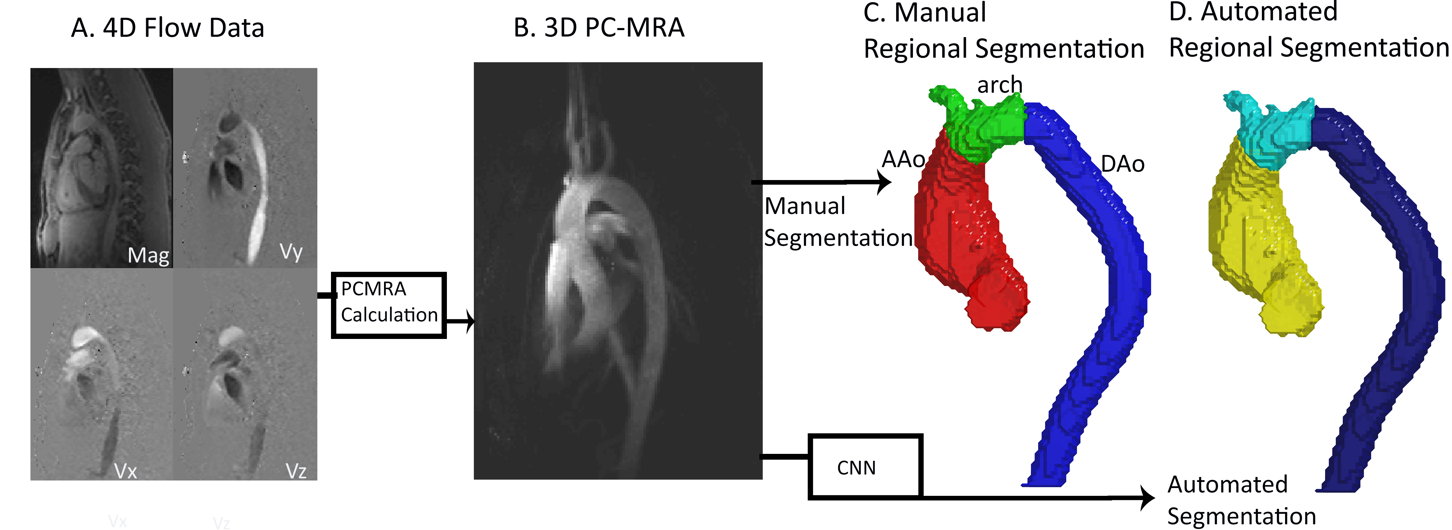
Figure
1: Workflow. All 4D flow data (Figure 1A) underwent standard 4D flow
preprocessing and used to generate 3D phase contract (PC) MRAs (Figure 1B). The
3D PCMRA was used to generate the ground-truth via manual or automated
segmentation of the aorta (utilizing a completely independent CNN) and manually
labeling the ascending aorta (AAo), arch, and descending aorta (DAo) (Figure
1C). The 3D PCMRA was, also, used as the input for the CNN, generating
automated segmentations (Figure 1D). Training and testing were performed
through 10-fold cross validation.
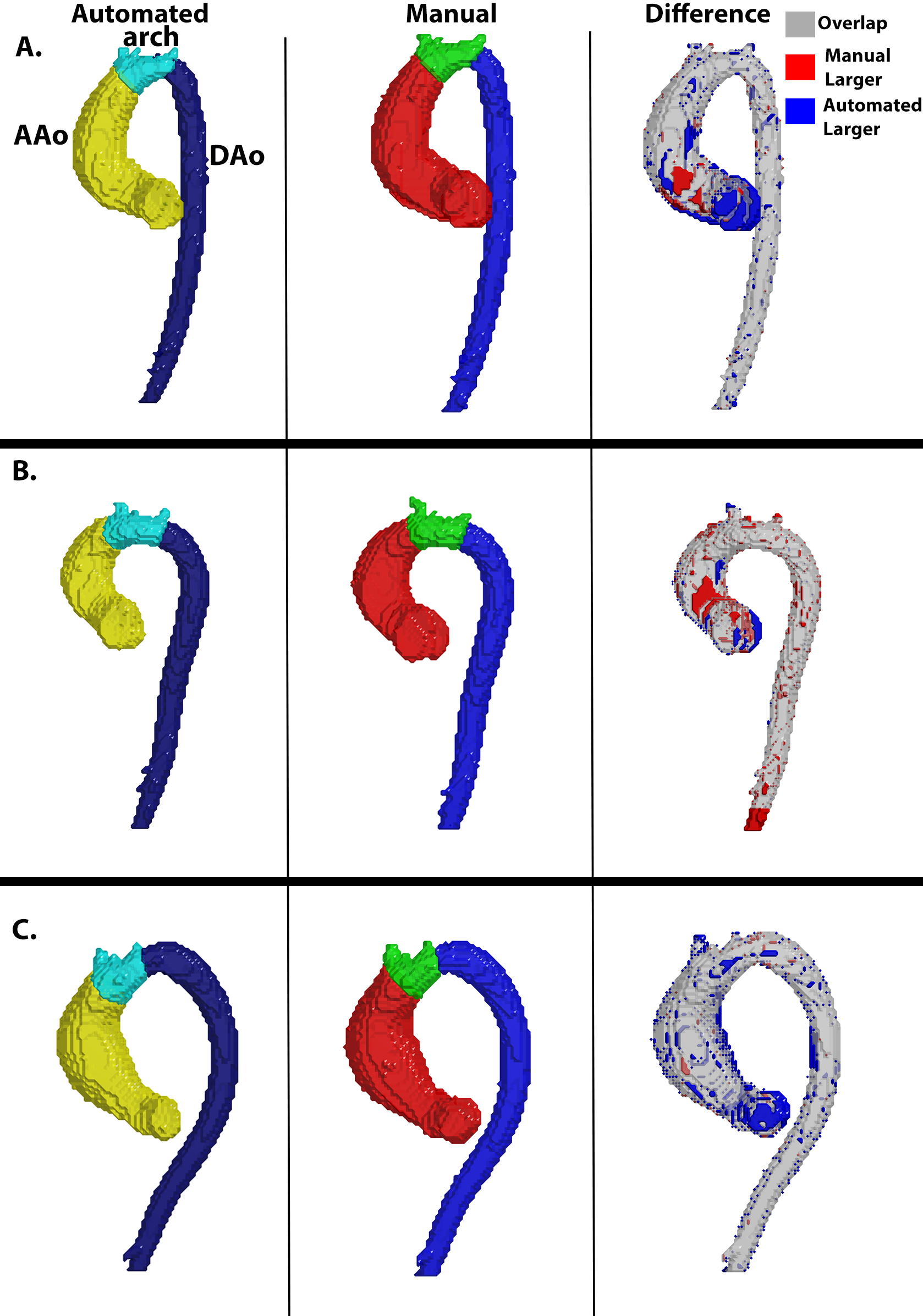
Figure
2: Examples of the Manual and Automated segmentations as well as a difference
map between them. Each example showcases a unique geometry of the aorta and
distinct placements of the aortic arch. In Figure 2A, the arch is located at
the peak of the aorta, while Figure 2B has a wider aortic arch, and for Figure
2C, the arch is slightly left of the top of the aorta. The Dice scores for
Figure 2A were AAo: 0.95, arch: 0.95, DAo: 0.98; for Figure 2B, AAo: 0.95,
arch: 0.88, DAo: 0.96; and Figure 2C, AAo: 0.95, arch: 0.86, DAo: 0.96