Pierre Daudé1, Patricia Ancel2, Sylviane Confort-gouny1, Anne Dutour2, Bénédicte Gaborit2, and Stanislas Rapacchi1
1Aix-Marseille Univ, CNRS, CRMBM, Marseille, France, 2APHM, Hôpital Universitaire Timone, Service d’Endocrinologie, Marseille, France
1Aix-Marseille Univ, CNRS, CRMBM, Marseille, France, 2APHM, Hôpital Universitaire Timone, Service d’Endocrinologie, Marseille, France
Deep-learning
segmentation of epicardial adipose tissue surface in 4CH cine proved the
evaluation of this long-overseen biomarker feasible in a database of 126. Networks
reached relative surface errors <20% within the upper half of the test set,
when 2 observers agreed within 15%.
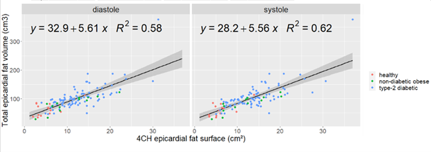
Figure 1 : Comparison of total epicardial fat volume
against 4-chamber surface measured on systolic or diastolic frame across the
three cohorts merged for the database.
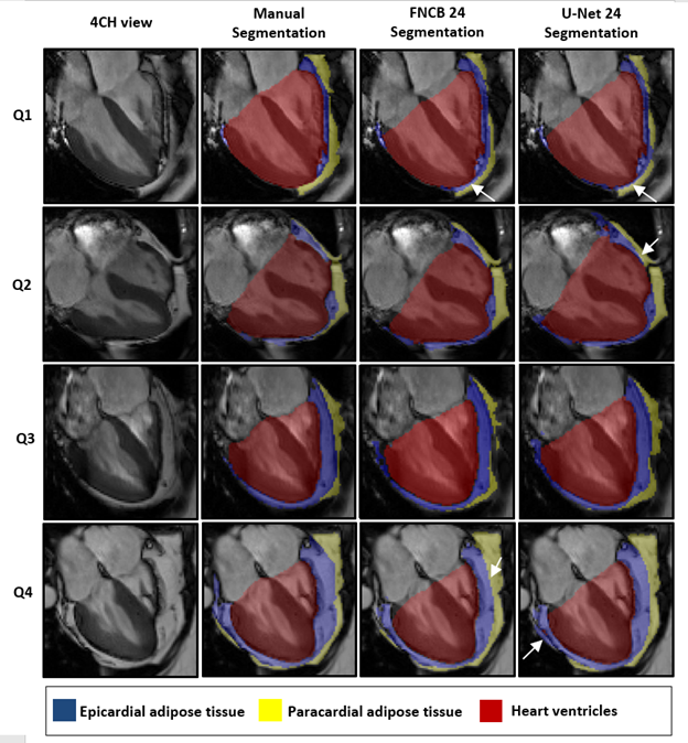
Figure 3 : Representative
automated segmentation results for each of EAT surface population quartile.
White arrows shows network segmentation errors.
