Matthew J. Middione1, Julio A. Oscanoa1,2, Michael Loecher1, Christopher M. Sandino3, Shreyas S. Vasanawala1, and Daniel B. Ennis1,4
1Department of Radiology, Stanford University, Palo Alto, CA, United States, 2Department of Bioengineering, Stanford University, Palo Alto, CA, United States, 3Department of Electrical Engineering, Stanford University, Palo Alto, CA, United States, 4Cardiovascular Institute, Stanford University, Stanford, CA, United States
1Department of Radiology, Stanford University, Palo Alto, CA, United States, 2Department of Bioengineering, Stanford University, Palo Alto, CA, United States, 3Department of Electrical Engineering, Stanford University, Palo Alto, CA, United States, 4Cardiovascular Institute, Stanford University, Stanford, CA, United States
In this work we analyzed the impact of reconstruction induced phase bias to determine the maximum acceleration factor that could be used with CS and DL reconstruction frameworks for 2D PC-MRI while minimizing errors in peak velocity and total flow within ±5%.
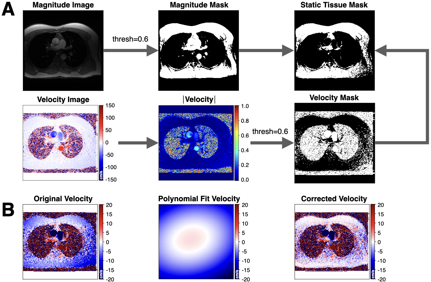
Figure 1. Overview of the background phase offset correction method. (A) Magnitude and velocity images were used as input to generate masked images of magnitude, velocity, and static tissue using a 60% signal intensity threshold. (B) The resulting static tissue mask was then used to generate a polynomial fit velocity image, which provides an estimated background phase offset image that can be used to correct the acquired velocity image.
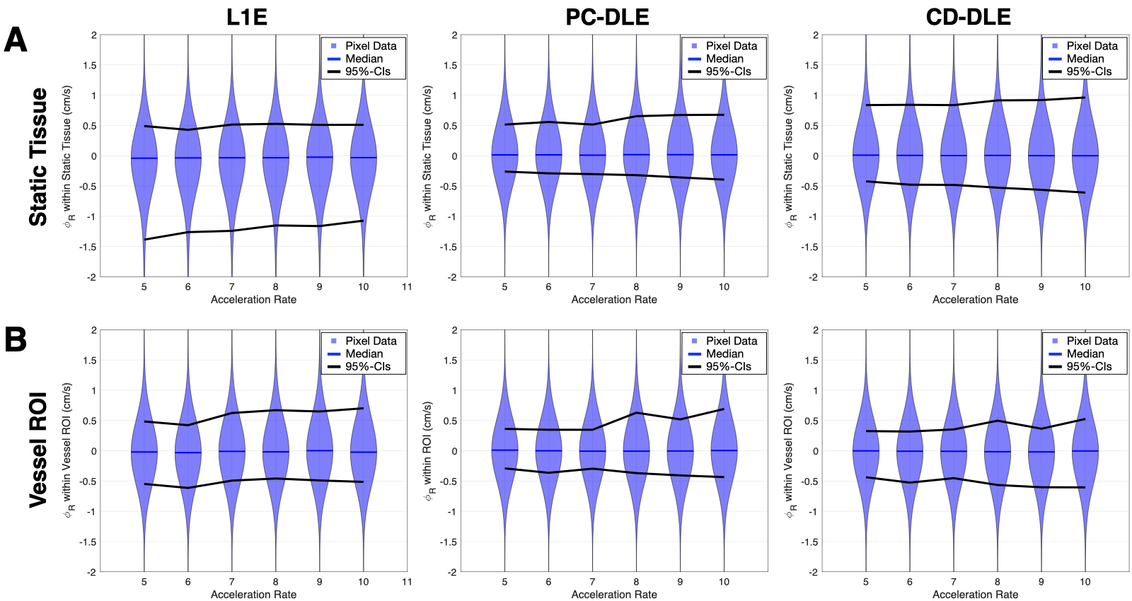
Figure 2. Pixel-by-pixel histogram differences demonstrate the magnitude of the reconstruction induced background phase offset bias, $$$\phi_{R}$$$ (cm/s), for L1E, PC-DLE and CD-DLE (A) within static tissue and (B) inside the vessel ROIs. The median (blue line) and 95%-CIs (lines) of $$$\phi_{R}$$$ are plotted as a function of the acceleration rate.