Ioannis Koktzoglou1,2, Rong Huang1, William J Ankenbrandt1,2, Matthew T Walker1,2, and Robert R Edelman1,3
1Radiology, NorthShore University HealthSystem, Evanston, IL, United States, 2University of Chicago Pritzker School of Medicine, Chicago, IL, United States, 3Northwestern University Feinberg School of Medicine, Chicago, IL, United States
1Radiology, NorthShore University HealthSystem, Evanston, IL, United States, 2University of Chicago Pritzker School of Medicine, Chicago, IL, United States, 3Northwestern University Feinberg School of Medicine, Chicago, IL, United States
DNN-based
SR reconstruction of 3D tsSOS-QISS MRA of the head and neck is feasible, and
potentially enables scan time reductions of 2-fold and 4-fold for portraying
the intracranial and extracranial arteries, respectively.
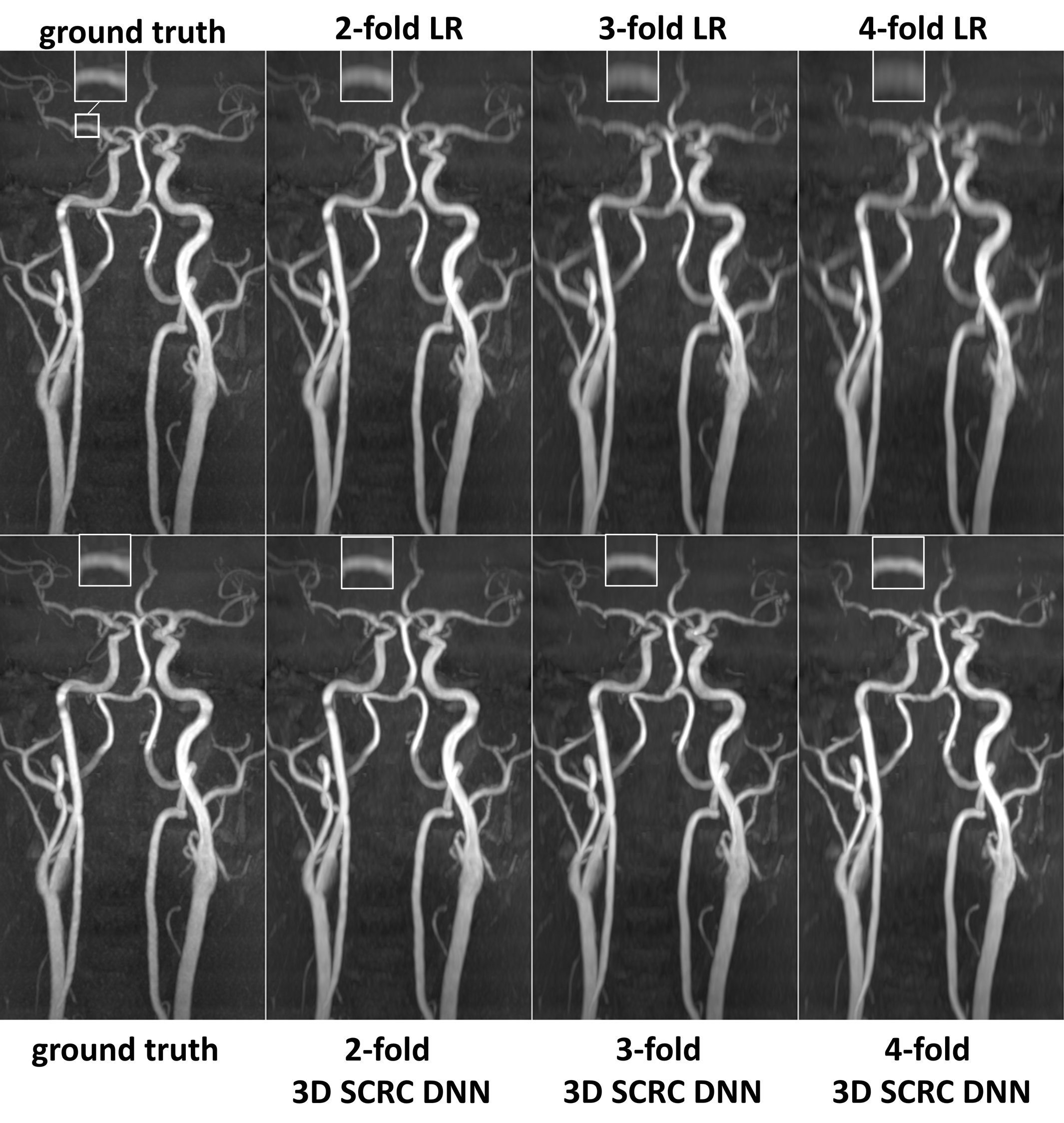
Figure 2. Coronal MIP 3D
tsSOS-QISS MRA images showing the impact of 3D SCRC SR DNN reconstruction on
image quality for 2- to 4-fold reduced of axial spatial resolution with respect
to ground truth data (left-most column) and input lower resolution (LR) data
(right-most upper panels). Insets show magnified views of the right middle
cerebral artery. Note the markedly improved image quality and spatial
resolution of the 3D SCRC SR DNN with respect to input LR volumes.
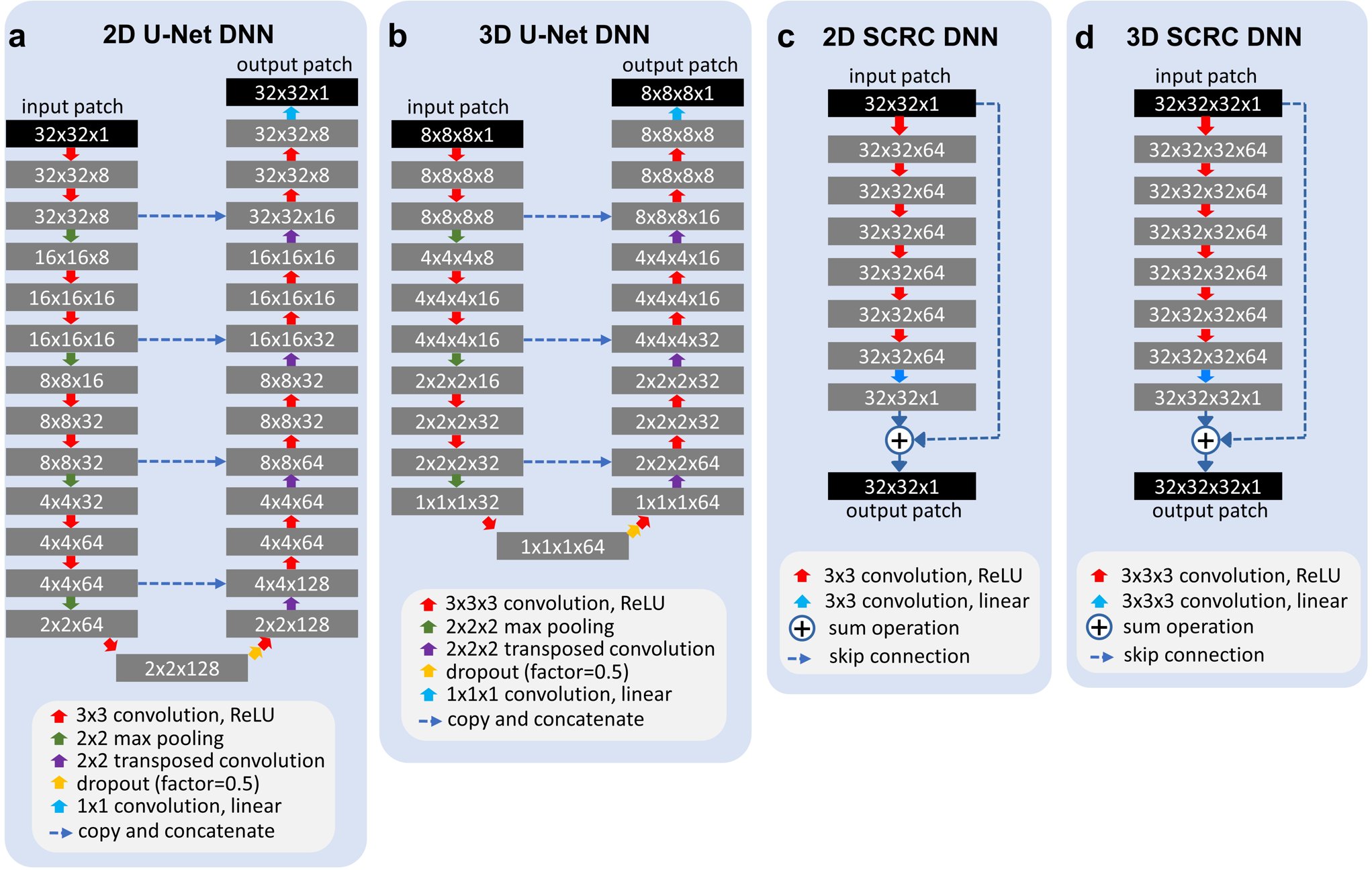
Figure 1. Architectures
of the deep neural networks used for super-resolution reconstruction. ReLU =
rectified linear unit. Training batch sizes for networks (a) through (d) were
400, 80, 400 and 20, whereas the number of trainable parameters for networks
were 540,073, 436,521, 185,857 and 556,801, respectively.