Nikou Louise Damestani1, David John Lythgoe1, Ana Beatriz Solana2, Brice Fernandez3, Steven Charles Rees Williams1, Fernando Zelaya1, and Florian Wiesinger2
1Department of Neuroimaging, King's College London, London, United Kingdom, 2ASL Europe, GE Healthcare, München, Germany, 3ASL Europe, GE Healthcare, Paris, France
1Department of Neuroimaging, King's College London, London, United Kingdom, 2ASL Europe, GE Healthcare, München, Germany, 3ASL Europe, GE Healthcare, Paris, France
We present a modification of a novel silent fMRI pulse sequence known as Looping Star. We show that this approach has additional benefits for high-resolution T2*-weighted structural imaging, quantitative susceptibility mapping and functional MRI.
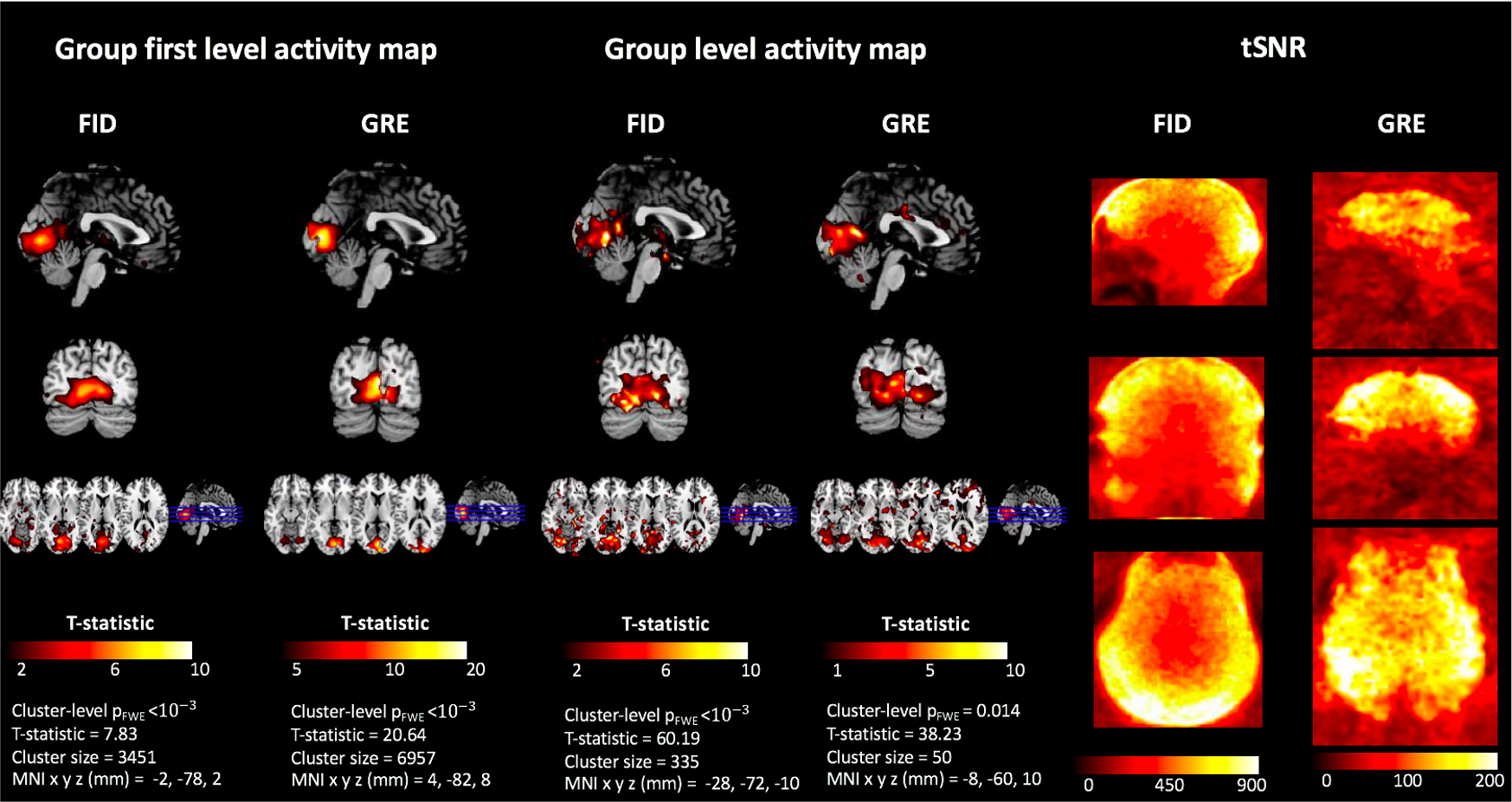
Figure 3: Output of fMRI analysis at group level and first level across participants for the FID and echo images. Significant (cluster level pFWE < 0.05) activity was identified in the visual cortex in both cases, with statistics listed below the figures. The tSNR maps of Looping Star are also illustrated on the right.
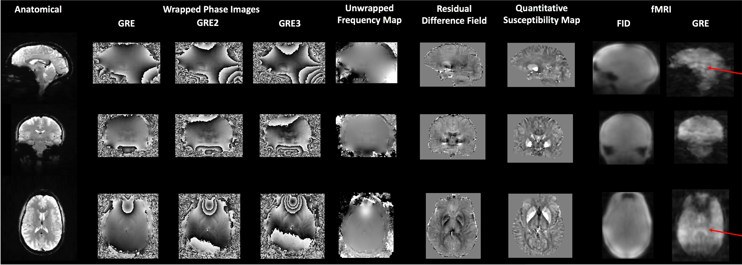
Figure 2: Output images from Looping Star acquisition techniques in a single participant. From left to right, the high resolution anatomical image, wrapped phase images, unwrapped frequency map, residual difference field, QSM maps and fMRI raw images are shown. Red arrows indicate clarity of ventricles in fMRI acquisition. QSM image range -0.1ppm to 0.1ppm.
