Kaibao Sun1, Zheng Zhong1,2, Muge Karaman1,2, Qingfei Luo1, and Xiaohong Joe Zhou1,2,3
1Center for MR Research, University of Illinois at Chicago, Chicago, IL, United States, 2Department of Bioengineering, University of Illinois at Chicago, Chicago, IL, United States, 3Departments of Radiology and Neurosurgery, University of Illinois at Chicago, Chicago, IL, United States
1Center for MR Research, University of Illinois at Chicago, Chicago, IL, United States, 2Department of Bioengineering, University of Illinois at Chicago, Chicago, IL, United States, 3Departments of Radiology and Neurosurgery, University of Illinois at Chicago, Chicago, IL, United States
A novel
simultaneous multi-segment (SMSeg) imaging method has been successfully applied
to fMRI for visualization of brain activations in multiple focal areas using
reduced FOVs with reduced geometric distortions.
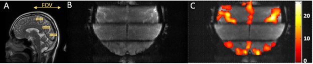
Figure
4: (A): The three segments at different locations as shown in yellow boxes
together with the FOV used in the acquisition. The lower and upper blocks covered
the visual cortex and motor cortex, respectively. (B): A composite SMSeg image
of (A) used in fMRI study (10 averages of images obtained during the baseline).
(C): The composite activation map from all three segments showed simultaneous
activations in the visual (the lower segment) and motor cortices (primary motor
cortex and supplementary motor area in the upper segment).
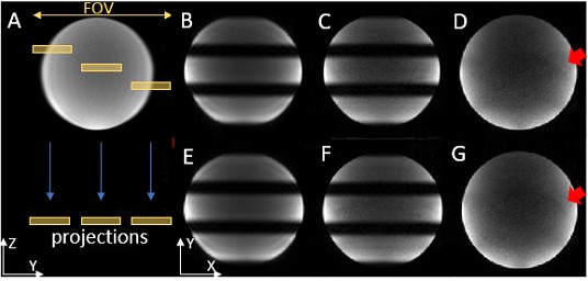
Figure
2: In (A), three obliquely distributed segments were excited simultaneously and
projected into one composite “slice”. Each column in the right panel shows two
representative axial composite slices of the phantom from the SMSeg EPI
sequence without in-plane acceleration (B, E) and with 4-fold acceleration (C,
F), together with the conventional 2D EPI with 4-fold acceleration at the
location corresponding to the central segment (D, G). The SNRs of images (C)
and (D) at the central segment were 50.9 and 46.3, respectively.