Rodolfo Abreu1, Miguel Castelo-Branco1,2, and João Valente Duarte1,2
1Coimbra Institute for Biomedical Imaging and Translational Research (CIBIT), Institute for Nuclear Sciences Applied to Health (ICNAS), University of Coimbra, Coimbra, Portugal, 2Faculty of Medicine, University of Coimbra, Coimbra, Portugal
1Coimbra Institute for Biomedical Imaging and Translational Research (CIBIT), Institute for Nuclear Sciences Applied to Health (ICNAS), University of Coimbra, Coimbra, Portugal, 2Faculty of Medicine, University of Coimbra, Coimbra, Portugal
Correction of geometric
distortions on fMRI data from phase-reversed images or field map images
improves the quality of different fMRI data analyses, with the latter yielding
the best results at the cost of longer scanning times.
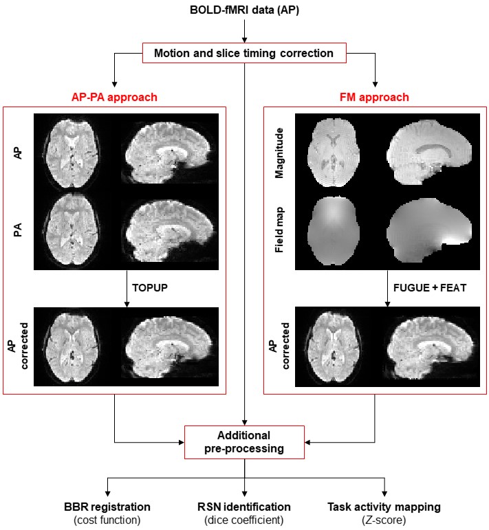
Fig. 1: Schematic diagram of the processing pipeline. The
fMRI data of each functional run is first submitted to motion and slice timing
correction. Then, geometric distortions are corrected using AP-PA or FM
approaches. Subsequently, additional pre-processing steps are performed on the
corrected and uncorrected data, followed by the three different data analyses: 1) registration into structural data, 2)
identification of resting-state networks, and 3) mapping of task-related brain
regions of interest.
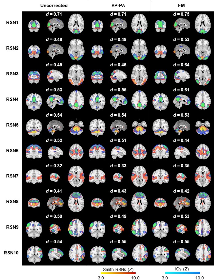
Fig. 3: Ten group RSNs
identified on (left) uncorrected, (middle) AP-PA corrected, and (right)
FM-corrected fMRI data for the first run of the biological motion
perception task. The RSN templates (in red-yellow) from 8 are superimposed with the spatial independent
components (in blue-light blue) selected for each RSN template, according to
their Dice coefficient (also shown above each RSN).