Michael Germuska1, Rachael Stickland2, Hannah Chandler3, and Richard Wise3,4
1School of Physics and Astronomy, Cardiff University, Cardiff, United Kingdom, 2Department of Physical Therapy and Human Movement Sciences, Northwestern University, Chicago, IL, United States, 3School of Psychology, Cardiff University, Cardiff, United Kingdom, 4Department of Neurosciences, University of Chieti-Pescara, Chieti, Italy
1School of Physics and Astronomy, Cardiff University, Cardiff, United Kingdom, 2Department of Physical Therapy and Human Movement Sciences, Northwestern University, Chicago, IL, United States, 3School of Psychology, Cardiff University, Cardiff, United Kingdom, 4Department of Neurosciences, University of Chieti-Pescara, Chieti, Italy
Monte-Carlo simulations show that resting cerebral metabolic rate of oxygen consumption can be estimated from resting perfusion (CBF) and the maximum BOLD signal (M). The method is demonstrated in-vivo using a repeated breath-holding protocol.
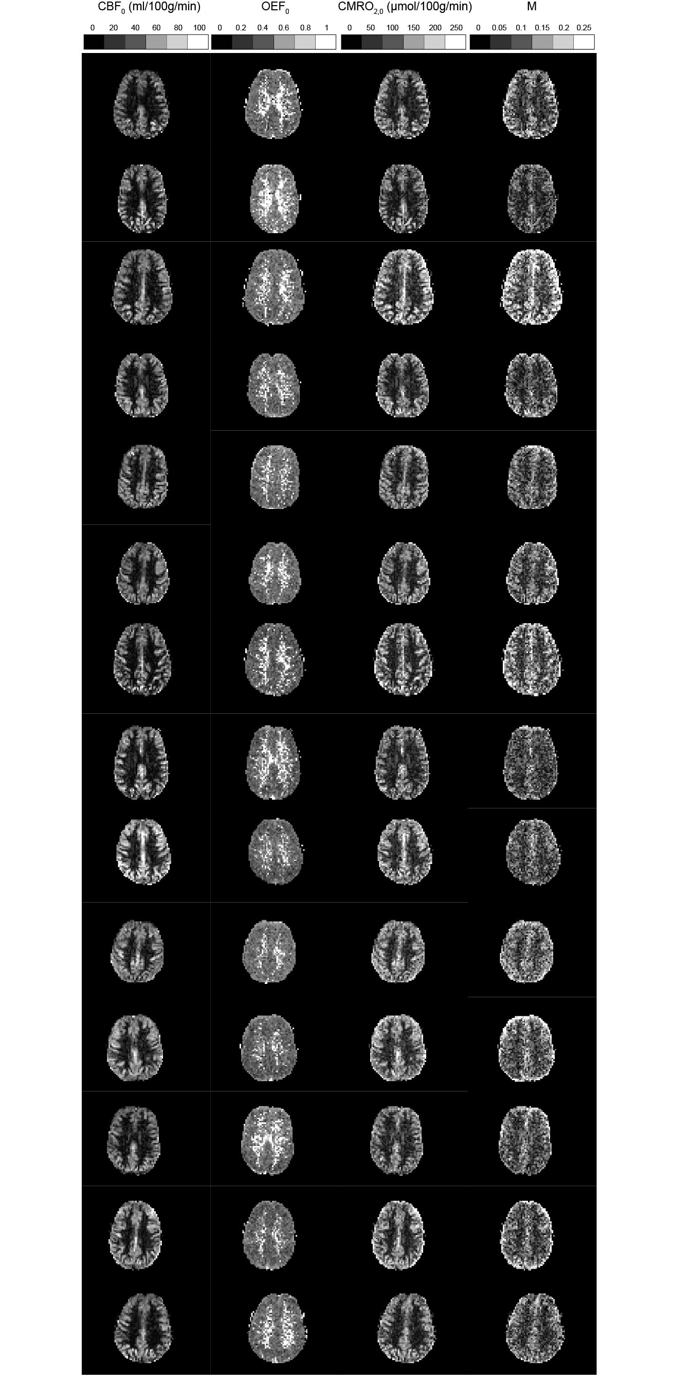
Figure 5. Example parameter maps for all subjects scanned. OEF estimates are approximately uniform in grey matter. High OEF estimates in the white matter are likely due to a lack of perfusion signal.