Myung-Ho In1, Daehun Kang1, Hang Joon Jo2, Uten Yarach3, Joshua D Trzasko1, Nolan K Meyer1,4, Bardwell Speltz J Lydia 1,4, John Huston III1, Matt A Bernstein1, and Yunhong Shu1
1Department of Radiology, Mayo Clinic, Rochester, MN, United States, 2Department of Physiology, College of Medicine, Hanyang University, Seoul, Korea, Republic of, 3Department of Radiologic Technology, Faculty of Associated Medical Sciences, Chiang Mai University, Chiang Mai, Thailand, 4Mayo Clinic Graduate School of Biomedical Sciences, Mayo Clinic, Rochester, MN, United States
1Department of Radiology, Mayo Clinic, Rochester, MN, United States, 2Department of Physiology, College of Medicine, Hanyang University, Seoul, Korea, Republic of, 3Department of Radiologic Technology, Faculty of Associated Medical Sciences, Chiang Mai University, Chiang Mai, Thailand, 4Mayo Clinic Graduate School of Biomedical Sciences, Mayo Clinic, Rochester, MN, United States
We performed breath-holding, reverse-gradient
fMRI with a PSF mapping-based distortion correction scheme and demonstrated the
effectiveness in improving fMRI reliability for each individual and the entire
group, especially in brain regions of rapid susceptibility change.
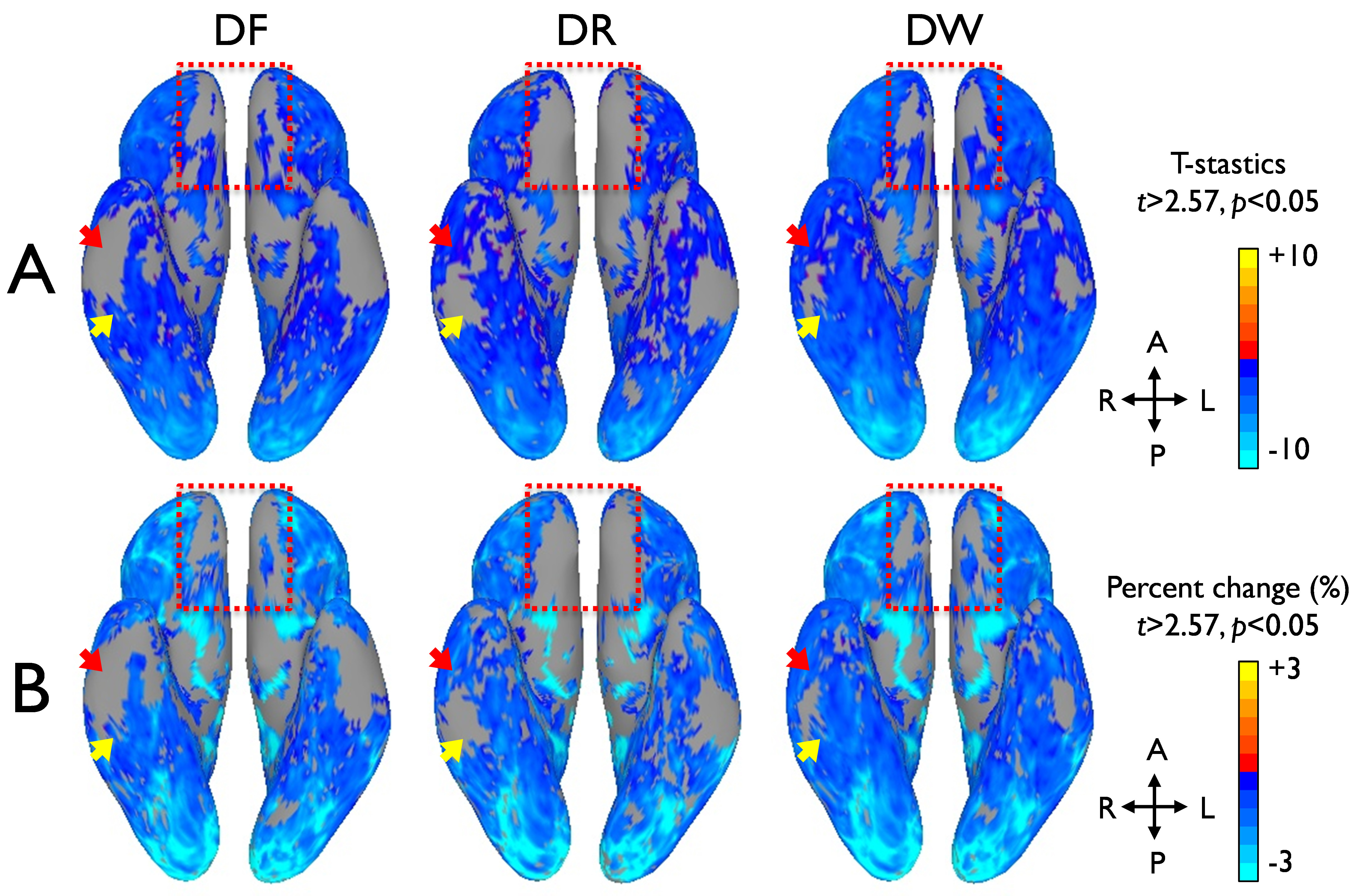
Figure 3. Comparison of group functional contrast maps between the
distortion-corrected forward (DF), the reverse (DR), and the combined EPI images
(DW) on a 3D inflated surface model. Group average maps of GLM T-statistics (A)
and signal percentage changes (B) are present for the comparison.
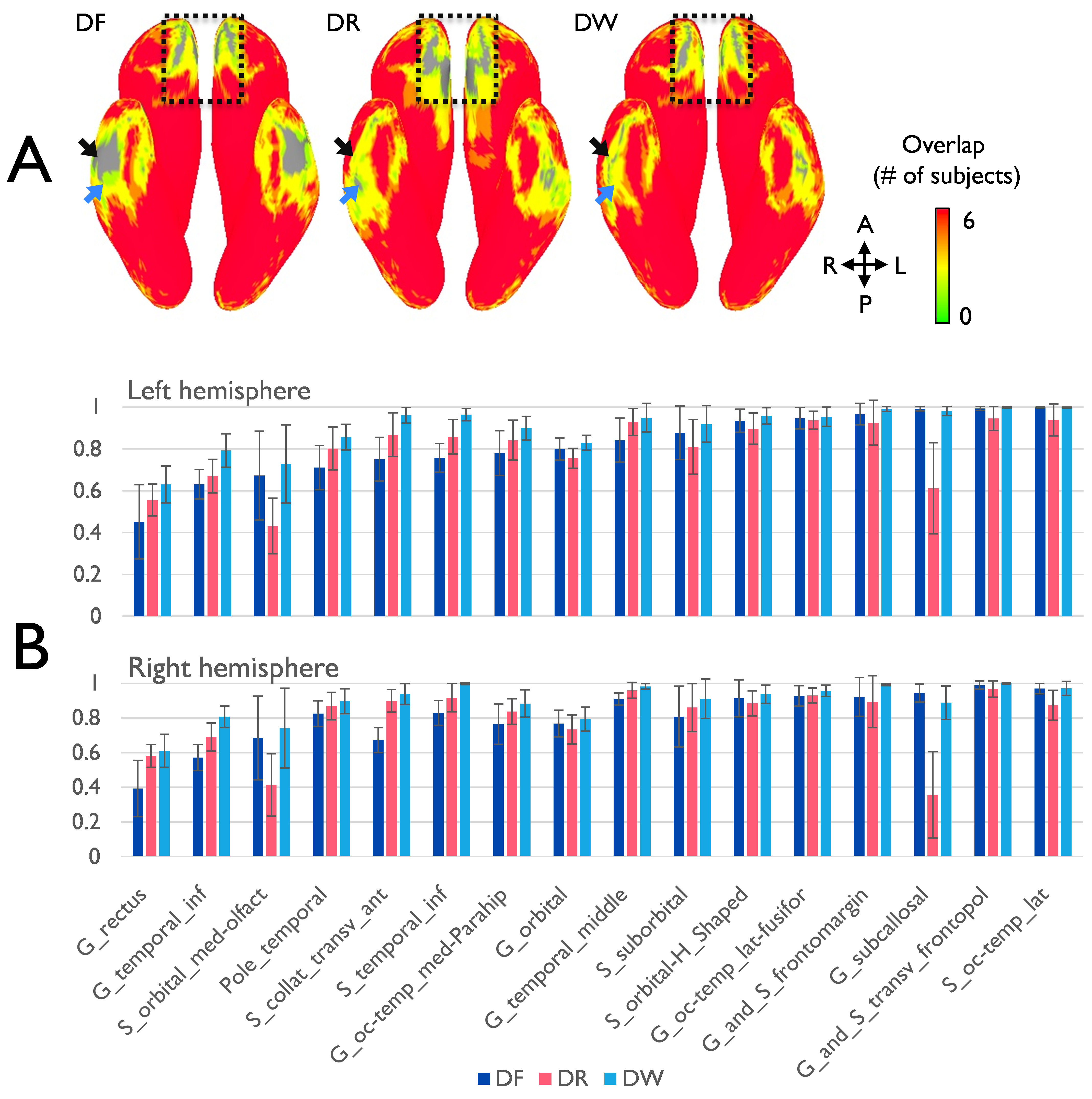
Figure 2. Comparison
of cortical coverage ratio between the distortion-corrected forward (DF), the
reverse (DR), and the combined EPI images (DW). (A) The cortical coverage for
six subjects is visualized on the inferior view of 3D inflated surface model.
(B) The coverage ratios at the local areas of both hemispheres are present as
the mean and standard deviation. The specific locations presenting apparent
coverage ratio differences are indicated by arrows and dashed-rectangular boxes
in (A).