Roberta Maria Lorenzi1, Alice Geminiani1, Claudia A.M. Gandini Wheeler-Kingshott1,2,3, Fulvia Palesi1,3, Claudia Casellato1, and Egidio D'Angelo1,3
1Department of Brain and Behavioral Sciences, University of Pavia, Pavia, Italy, 2NMR Research Unit, Queen Square MS Centre, Department of Neuroinflammation, UCL Queen Square Institute of Neurology, Faculty of Brain Sciences, University College London (UCL), London, United Kingdom, 3Brain Connectivity Centre Research Department, IRCCS Mondino Foundation, Pavia, Italy
1Department of Brain and Behavioral Sciences, University of Pavia, Pavia, Italy, 2NMR Research Unit, Queen Square MS Centre, Department of Neuroinflammation, UCL Queen Square Institute of Neurology, Faculty of Brain Sciences, University College London (UCL), London, United Kingdom, 3Brain Connectivity Centre Research Department, IRCCS Mondino Foundation, Pavia, Italy
Cerebellum physiological-grounded mean-field model was defined as a set of equations modelling the output activity of each neural populations. Transfer function fitting pipeline was tested at different spiking-rate input showing populations parameters reliable trend.
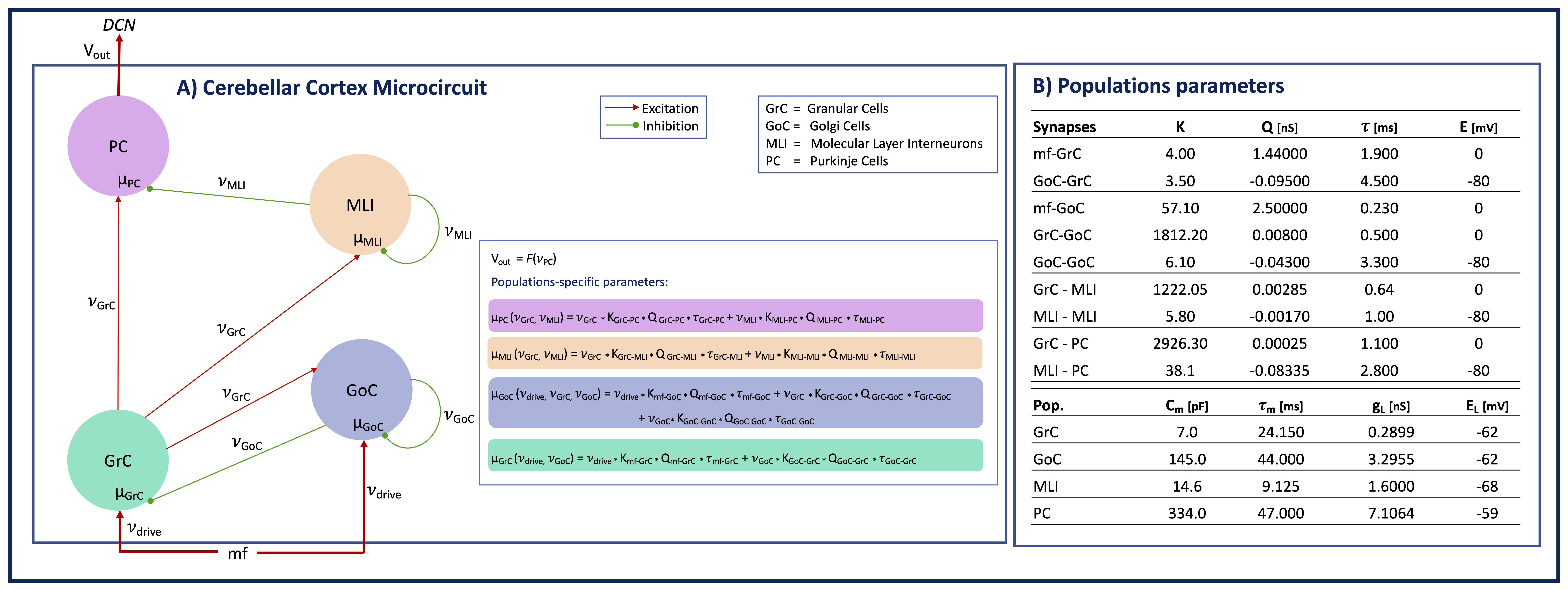
Figure 1) Cerebellar cortex microcircuit model. Populations (p) = GrC, GoC, MLI, PC. Connections (c) = mf-GrC, mf-GoC, GrC-GoC, GoC-GrC, GoC-GoC, GrC-MLI, MLI-MLI, GrC-PC, MLI-PC. The external input 𝜈input[Hz] is relayed by mossy fibers (mf) to GrC and GoC. The output activity 𝜈PC projects to the Deep Cerebellar Nuclei (DCN). Per p or c: mp=conductance; 𝜈p=firing rate[Hz]; Kc=connections probability*cells numbers; Qc = quantal synaptic conductance, 𝜏c=synaptic time decay.
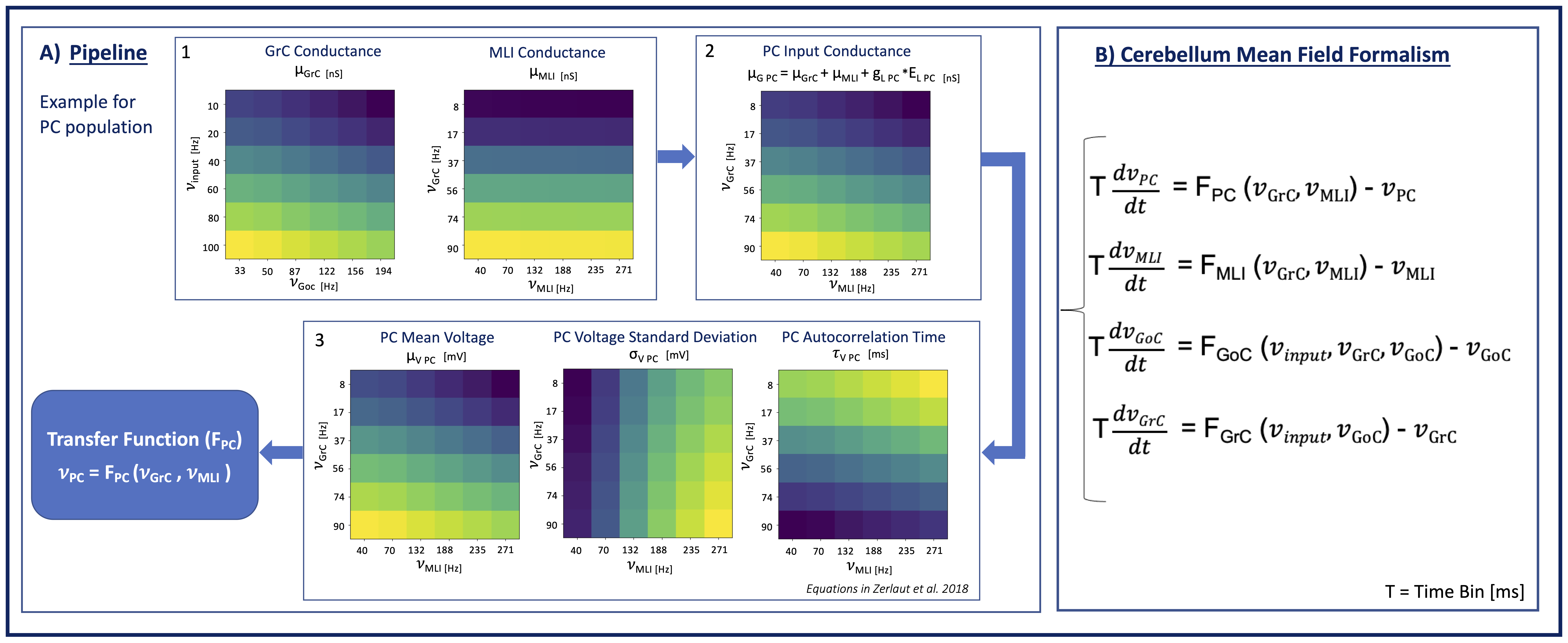
Figure 2) Pipeline to compute the transfer function (F). A): Color-map showing from max(yellow) to min(blue) parameters values used for F fitting. A1) Population conductances assume high values for high excitation combined with low inhibition. A2) PC input conductance depending on connected populations conductances. A3) PC membrane voltage properties: μV follows the same conductances trend. σV is higher for low excitation and high inhibition. 𝜏V is higher for low excitation-high inhibition. B): Cerebellum Mean-Field Model describing activity evolution of each populations