Seon-Ha Hwang1, SoHyun Han2, Seong-Gi Kim2, Jaeseok Park3,4, and Sung-Hong Park1
1Department of Bio and Brain Engineering, Korea Advanced Institute of Science and Technology, Daejeon, Korea, Republic of, 2Center for Neuroscience Imaging Research, Institute of Basic Science, Suwon, Korea, Republic of, 3Department of Biomedical Engineering, Sungkyunkwan University, Suwon, Korea, Republic of, 4Department of Intelligent Precision Healthcare Convergence, Suwon, Korea, Republic of
1Department of Bio and Brain Engineering, Korea Advanced Institute of Science and Technology, Daejeon, Korea, Republic of, 2Center for Neuroscience Imaging Research, Institute of Basic Science, Suwon, Korea, Republic of, 3Department of Biomedical Engineering, Sungkyunkwan University, Suwon, Korea, Republic of, 4Department of Intelligent Precision Healthcare Convergence, Suwon, Korea, Republic of
The proposed SAR-efficient non-segmented 3D-EPI-pCASL produced high-quality perfusion maps at both 3T and 7T. Its SAR efficiency and high temporal resolution enabled us to get whole-brain 3D-pCASL fMRI map at 7T for the first time.
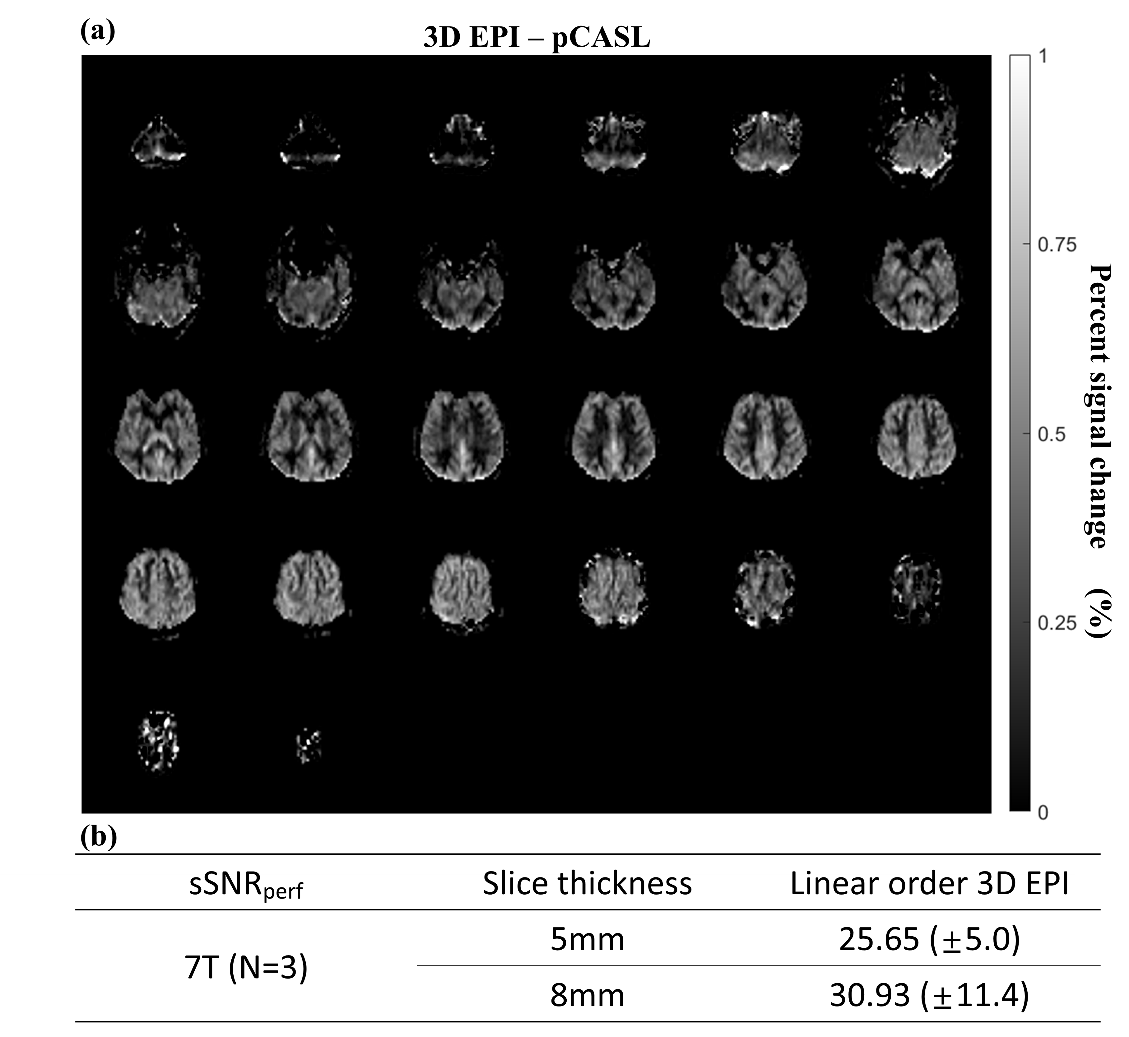
Fig. 3. Representative percent signal
change map of 3D EPI-pCASL and
spatial SNR of perfusion map at 7T. (a) CBF map by 3D EPI-pCASL with
5mm slice thickness. (b) Table of spatial SNR of perfusion map by thickness.
Despite limitation of coil coverage and B0, B1 inhomogeneity, the perfusion map
from 7T MRI have consistent quality with that of 3T. Also, spatial SNR is
similar with that of 3T. Because lowered
labeling efficiency by hardware limitation could not be calculated, percent
signal change map is represented instead of CBF map.
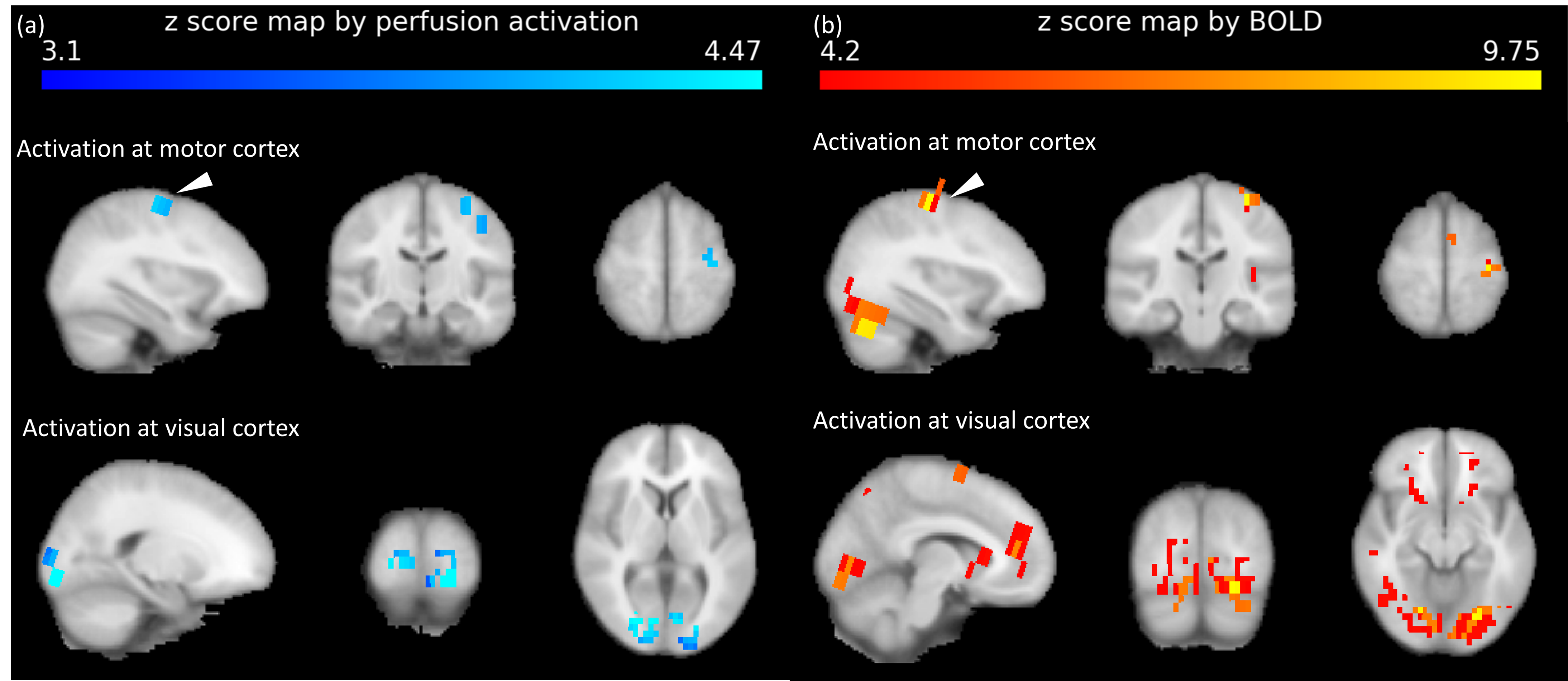
Fig. 4. Functional activation map by
(a)perfusion and (b)BOLD activation. For the both perfusion and BOLD,
activations were shown simultaneously at visual (occipital lobe) and motor
cortex (white arrows). Although the sensitivity of perfusion activation is
lower than that of BOLD, spatial localization is much specified by perfusion
activation map.