Gael Saib1, Alan Koretsky1, and S Lalith Talagala2
1NINDS/LFMI, National Institutes of Health, Bethesda, MD, United States, 2NINDS/NMRF, National Institutes of Health, Bethesda, MD, United States
1NINDS/LFMI, National Institutes of Health, Bethesda, MD, United States, 2NINDS/NMRF, National Institutes of Health, Bethesda, MD, United States
The maximal labeling efficiency of PCASL at 7T was experimentally measured at ~0.6 using a 1Tx/32Rx head coil with a PCASL-prepared FLASH sequence.

Figure 2: PCASL labeling efficiency as a function of B1mean measured in ICAs with Gmean 0.25mT/m. For 4 of the arteries, the labeling efficiency maximized ~0.6 at a B1mean of ~0.4uT. Note that the maximum achievable B1mean was limited in RICA of subject 3 compared to others due to increased B1+ inhomogeneities in the labeling plane.
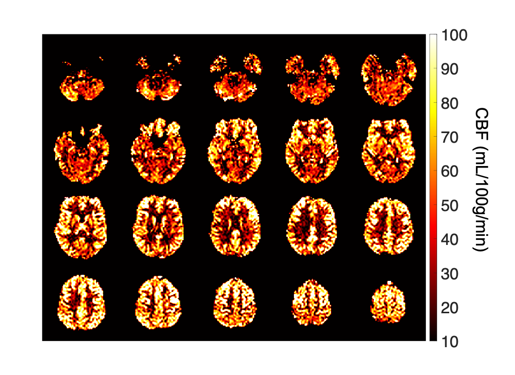
Figure 4: Whole-brain CBF maps acquired with a 2D-EPI PCASL sequence at 7T. The PCASL sequence parameters were Gmax=3.5mT/m, Gmean=0.25mT/m, Hanning RF pulse duration/separation=800/1800s, nominal FA=35°, labeling duration/PLD=1.5s, matrix=64x64, resolution=3x3x3mm3, 23 slices, TE/TR=12/4500ms, GRAPPA factor=2 and TA=6min. With a labeling efficiency of 0.6, the average CBF in the cortex was about 66mL/100g/min.