Peiyao Chen1, Chao Jin1, Xianjun Li1, Miaomiao Wang1, Congcong Liu1, Xiaoyu Wang1, Fan Wu1, Yuli Zhang1, Cong Tian1, Mengxuan Li1, Xiaocheng Wei2, and Jian Yang1
1First Affiliated Hospital of Xi 'an Jiaotong University, Xi'an, Shaanxi, China, 2MR Research China, GE Healthcare, Beijing, China
1First Affiliated Hospital of Xi 'an Jiaotong University, Xi'an, Shaanxi, China, 2MR Research China, GE Healthcare, Beijing, China
The
estimated age with highest cerebral blood flow is earliest in the occipital
lobe, followed by temporal and parietal lobe, at last in the frontal lobe. The
perfusion of basal ganglia shows a U-shaped curve, which slowly increases with
age.
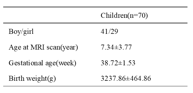
Table 1 Demographic
data Note: Mean±SD
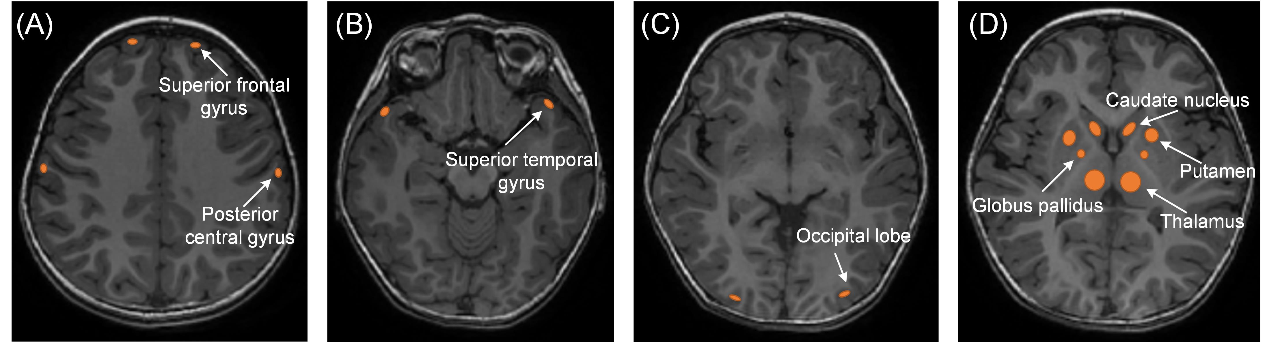
Figure
1. Manual regions of interest(ROIs)were placed on the CBF map by using the
aligned anatomical image as guidance. ROI was about 20-100mm2. (A)bilateral
superior frontal gyrus and posterior central gyrus .(B)bilateral superior
temporal gyrus.(C)bilateral occipital lobe(D)basal ganglia (bilateral thalamus,
globus pallidus, putamen, caudate nucleus )