Hongri Chen1, Weiqiang Dou2, and Wei yin Liu2
1Dalian Medical University, Northern Jiangsu People’s Hospital, Yangzhou, China, Yangzhou, China, 2GE Healthcare, MR Research China, Beijing, P.R. China, Beijing, China
1Dalian Medical University, Northern Jiangsu People’s Hospital, Yangzhou, China, Yangzhou, China, 2GE Healthcare, MR Research China, Beijing, P.R. China, Beijing, China
The normalized cerebral blood flow (CBF) provided sensitive imaging-based markers that contribute to the differential diagnosis of the Parkinson's disease dementia (PDD) and Alzheimer’s disease (AD).
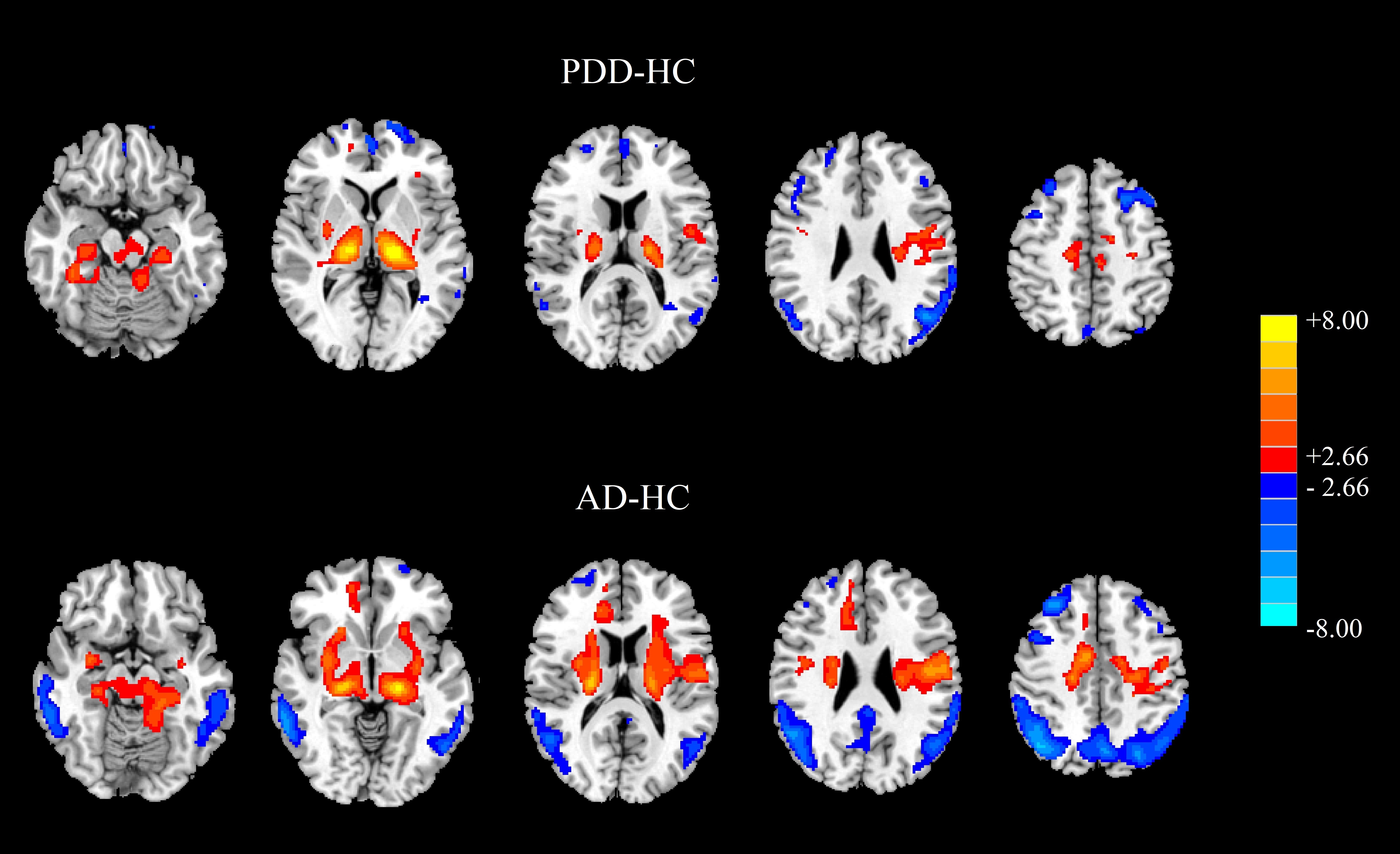
Figure 1 Results of two‐sample t‐test between dementia
patients and HC subjects. The above: PDD minus HC. The below: AD minus HC. Red color
represents the increased perfusion, while the blue color represents the
decreased perfusion (PFWE < 0.001).
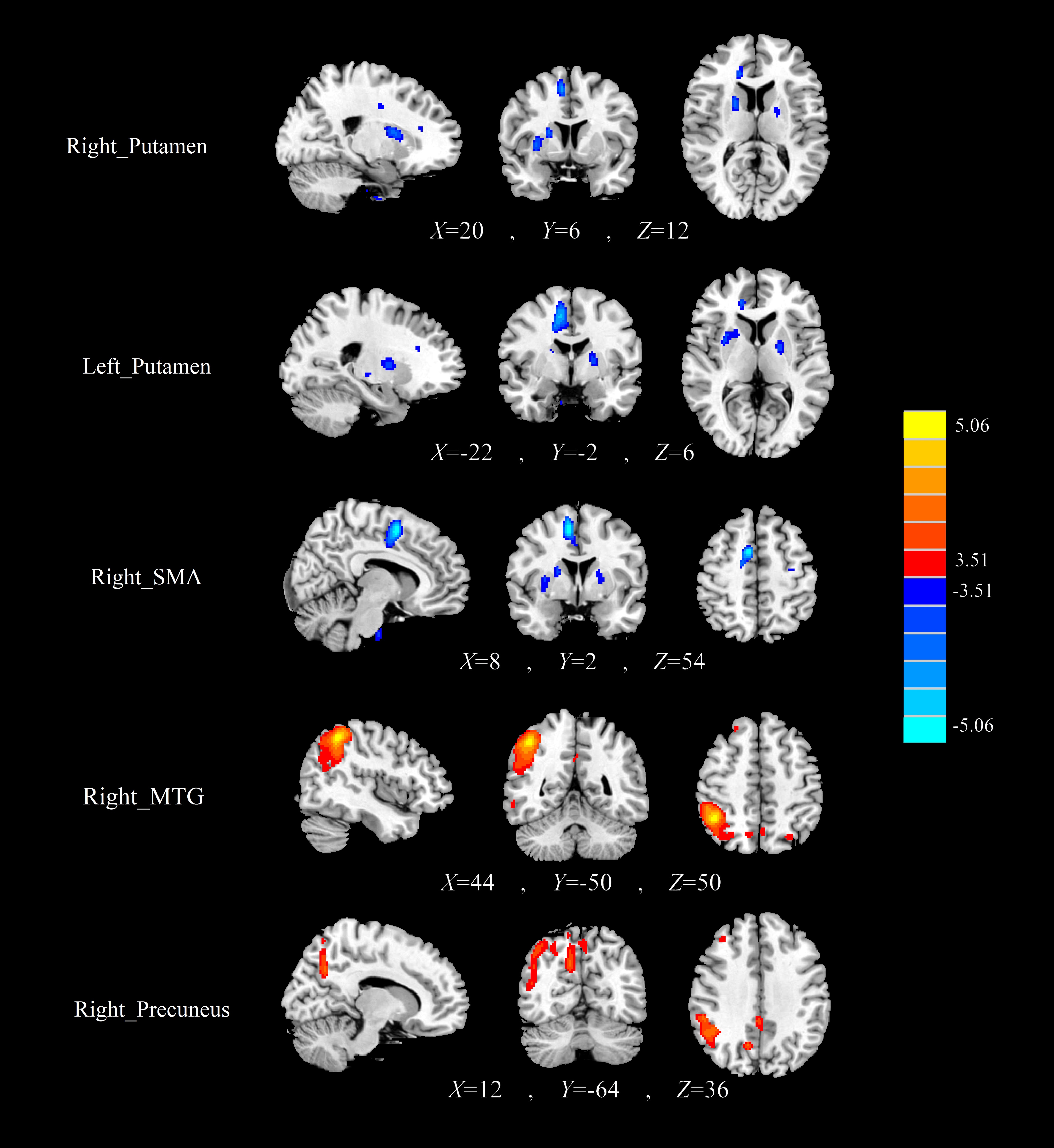
Figure
2 The normalized CBF differences between the PDD patients and the AD patients.
Compared with AD, the PDD patients showed decreased CBF in the bilateral putamen
and right SMA, as well as increased CBF in the right MTG, and right precuneus.