Sudipto Dolui1, Audrey P. Fan2,3, Moss Y. Zhao3, Greg Zaharchuk3, and John A. Detre1,4
1Department of Radiology, University of Pennsylvania, Philadelphia, PA, United States, 2Departments of Biomedical Engineering and Neurology, University of California, Davis, CA, United States, 3Department of Radiology, Stanford University, Palo Alto, CA, United States, 4Department of Neurology, University of Pennsylvania, Philadelphia, PA, United States
1Department of Radiology, University of Pennsylvania, Philadelphia, PA, United States, 2Departments of Biomedical Engineering and Neurology, University of California, Davis, CA, United States, 3Department of Radiology, Stanford University, Palo Alto, CA, United States, 4Department of Neurology, University of Pennsylvania, Philadelphia, PA, United States
Cerebral blood flow in periventricular white matter measured using state-of-the-art arterial spin labeling showed high intra-session reproducibility and correlated with measurements from concurrently acquired [15O]-water PET, demonstrating it can be measured reliably.
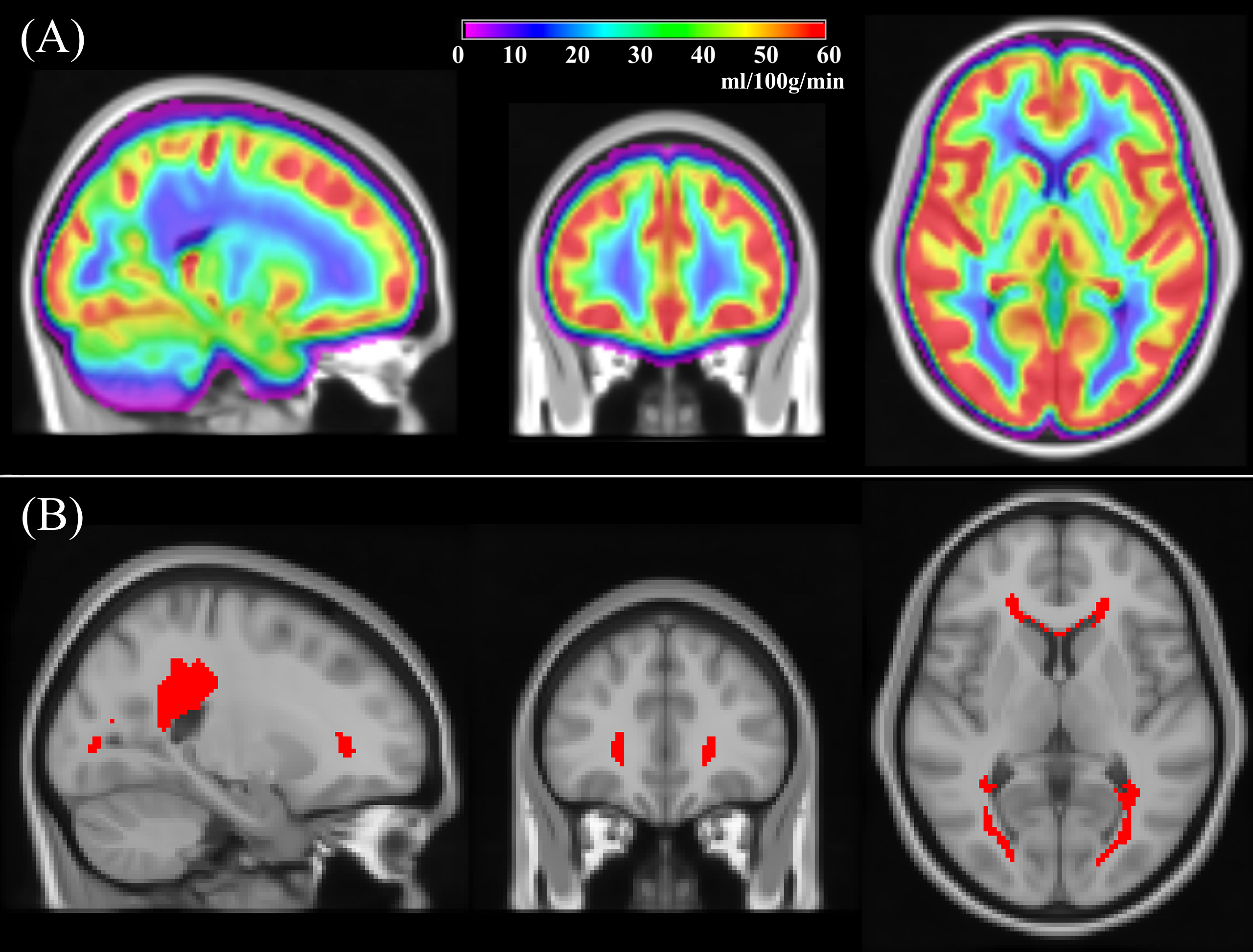
Figure 1: (A) Group averaged CBF obtained from 436 subjects (age=50.4±3.5 years, 54% female) from the NHLBI Coronary Artery Risk Development in Young Adults (CARDIA) study; (B) A periventricular white matter (PVWM) region of interest (ROI) obtained by thresholding the group averaged map in (A) to CBF<12.5ml/100g/min.
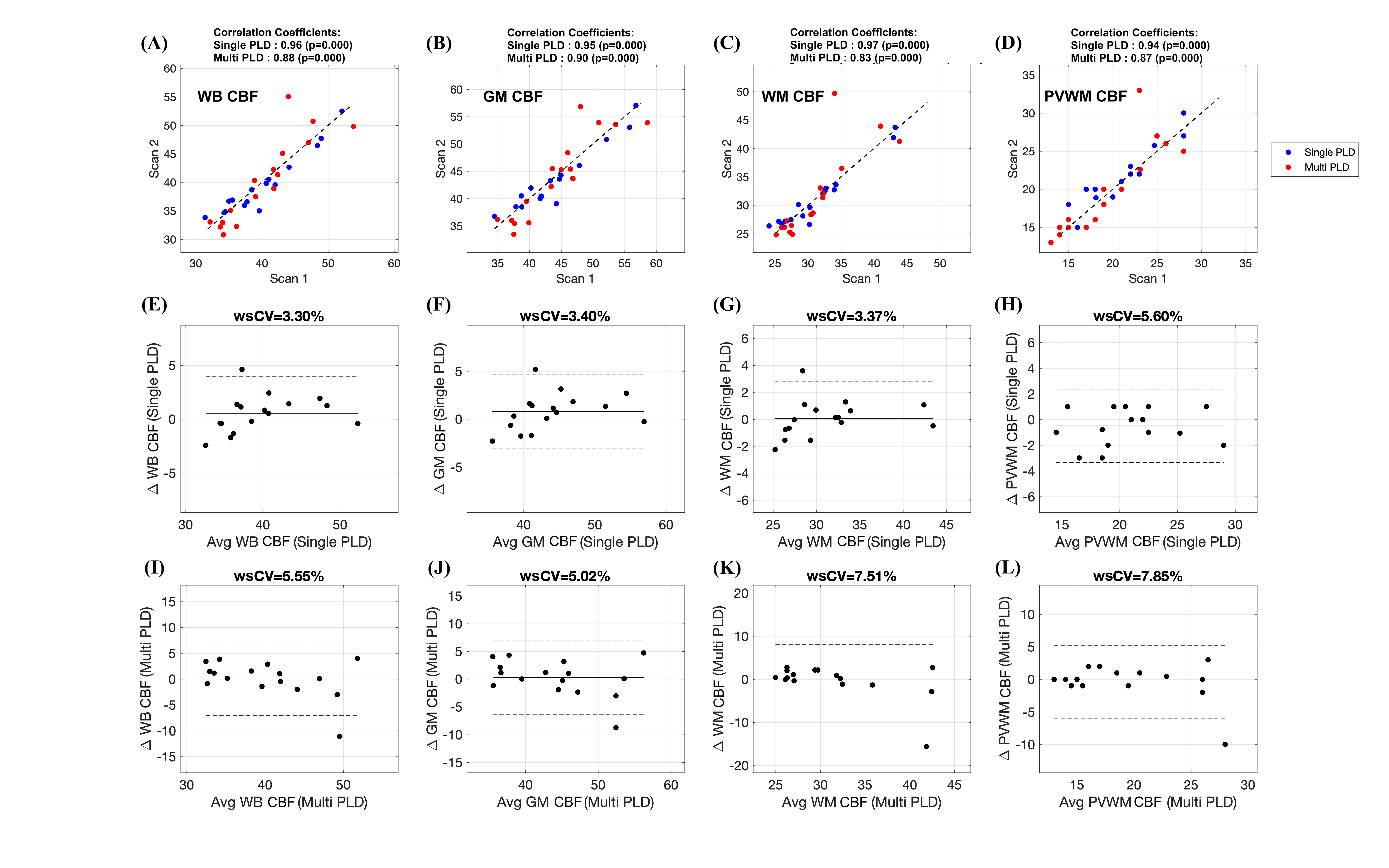
Figure 2: (A)-(D): Scatter plots comparing the (A) whole brain (WB), (B) gray matter (GM), (C) white matter (WM) and (D) periventricular WM (PVWM) CBF obtained from the two sessions using single-PLD (shown in blue) and multi-PLD (shown in red) ASL. The black lines correspond to the unity line and the titles show the correlation coefficients. (E)-(H) and (I)-(L) show Bland-Altman Plots corresponding to (E & I) WB CBF, (F & J) GM CBF, (G & K) WM CBF and (H & L) PVWM CBF using the two sessions of single and multi-PLD data respectively. The wsCV values are in the titles of the plots.