Bhaswati Roy1, Susana Vacas1, Kathy McCloy2, Rajan Saggar2, and Rajesh Kumar1,3,4,5
1Anesthesiology, University of California Los Angeles, Los Angeles, CA, United States, 2Medicine, University of California Los Angeles, Los Angeles, CA, United States, 3Bioengineering, University of California Los Angeles, Los Angeles, CA, United States, 4Radiological Sciences, University of California Los Angeles, Los Angeles, CA, United States, 5Brain Research Institute, University of California Los Angeles, Los Angeles, CA, United States
1Anesthesiology, University of California Los Angeles, Los Angeles, CA, United States, 2Medicine, University of California Los Angeles, Los Angeles, CA, United States, 3Bioengineering, University of California Los Angeles, Los Angeles, CA, United States, 4Radiological Sciences, University of California Los Angeles, Los Angeles, CA, United States, 5Brain Research Institute, University of California Los Angeles, Los Angeles, CA, United States
PAH
patients show cognitive and mood deficits, and brain changes in those sites. However,
the underlying cause of tissue damage in PAH patients remain unclear. We show
reduced regional CBF in PAH patients over controls, and correlations between CBF
and functional deficits in the condition.
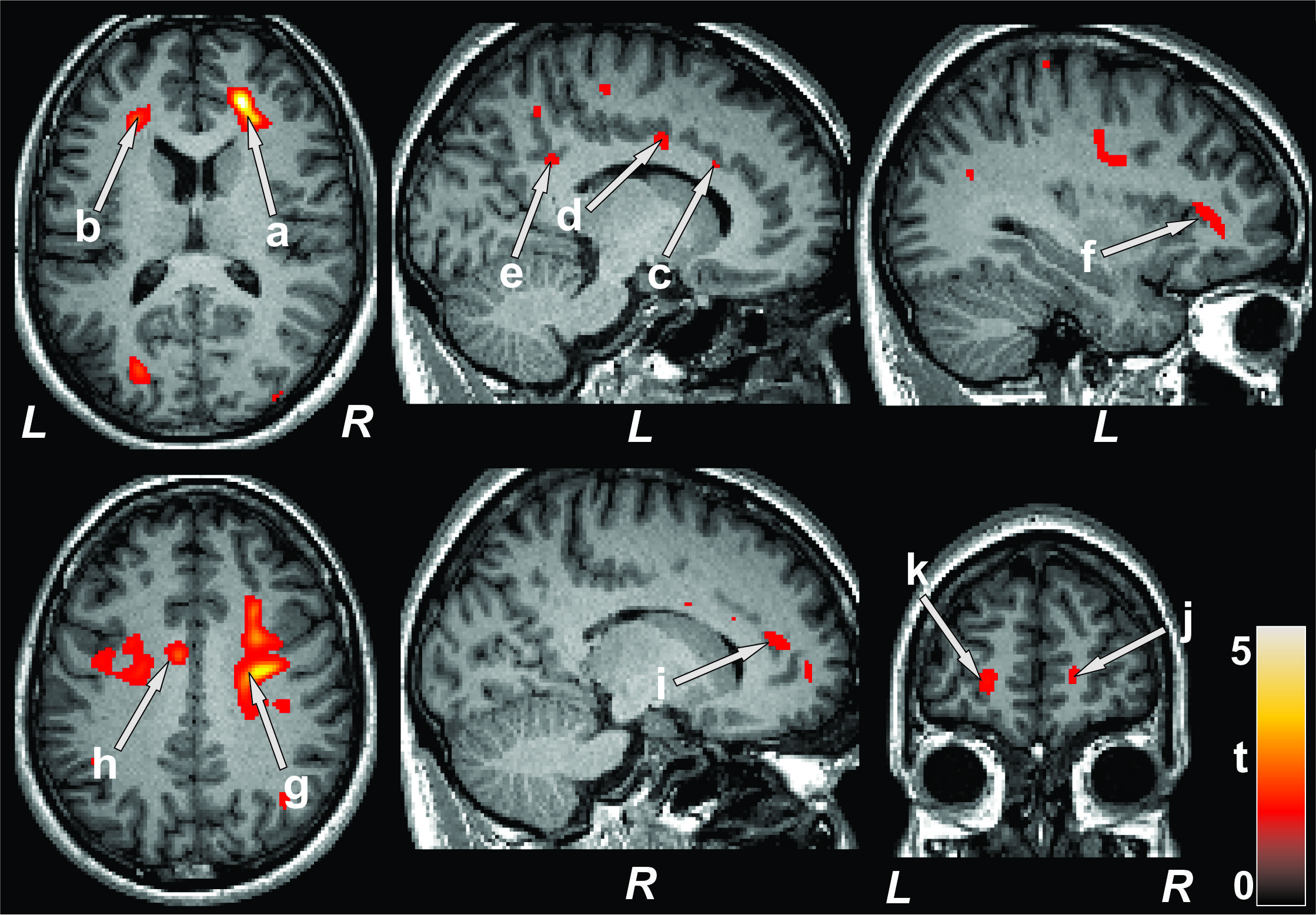
Figure 1: Brain regions with reduced CBF in PAH patients over
control subjects. The sites with reduced CBF in PAH patients included the
bilateral frontal white matter (a, b), left anterior (c), mid (d), and
posterior (e), and right anterior (i) cingulate, left insula (f), bilateral corona
radiata (g, h), and bilateral prefrontal cortices (j, k). All images are in
neurological convention (L = left; R = right). Color bar indicates t-statistic
values.
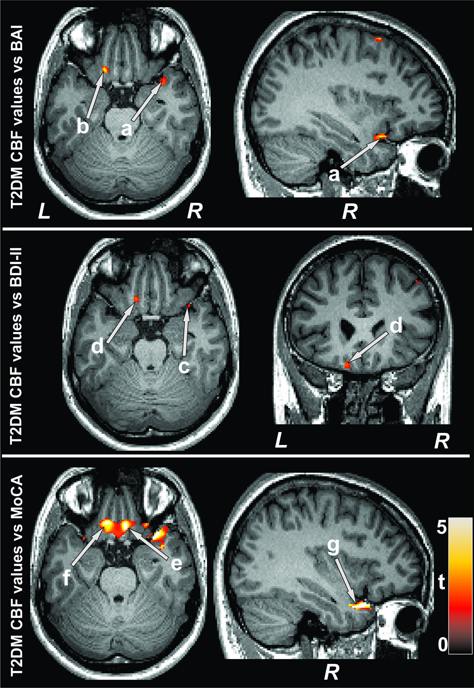
Figure 2: Negative correlations emerged between CBF and mood symptoms
and positive associations between cognition and CBF in PAH patients. Correlations
appeared between mood scores and CBF values of the right insula (a, c), and left
basal forebrain (b, d), and between MoCA scores and CBF values of the bilateral
basal forebrain (e, f) and right insula (g). Figure conventions are same as in
Figure 1.