Colette C. Milbourn1, Thomas W. Okell2, and Nicholas P. Blockley1
1The School of Life Sciences, University of Nottingham, Nottingham, United Kingdom, 2Wellcome Centre for Integrative Neuroimaging, FMRIB, Nuffield Department of Clinical Neurosciences, University of Oxford, Oxford, United Kingdom
1The School of Life Sciences, University of Nottingham, Nottingham, United Kingdom, 2Wellcome Centre for Integrative Neuroimaging, FMRIB, Nuffield Department of Clinical Neurosciences, University of Oxford, Oxford, United Kingdom
Cerebrovascular reactivity measured with Pseudocontinuous Arterial Spin Labelling (pCASL) systematically varies with different pCASL preparation parameters due to changes in blood velocity in the internal carotid and vertebral arteries.
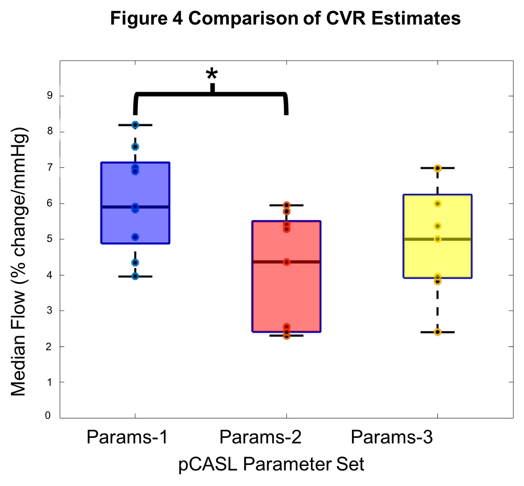
Figure 4 Comparison of CVR estimates. Box plots shows the median cerebrovascular reactivity (CVR) for each of the three pCASL (pseudocontinuous Arterial Spin Labelling) Parameter sets. CVR is the percentage change in cerebral blood flow per mmHg partial pressure of end-tidal carbon dioxide (PetCO2).
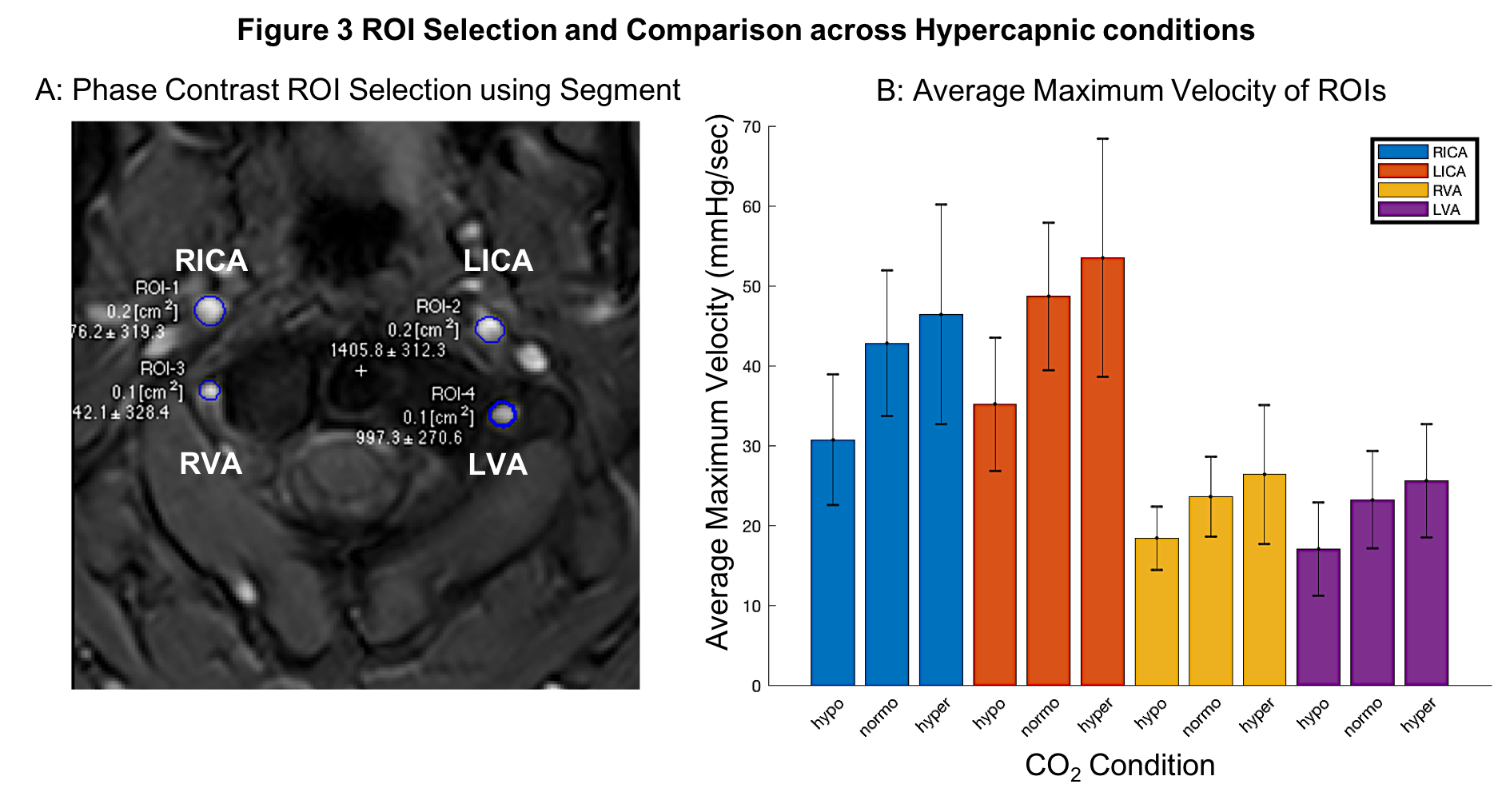
Figure 3 ROI Selection and Blood Velocity Results. A: Phase contrast velocity region of interest (ROI) selection of left/right (L/R) internal carotid arteries (ICA) and vertebral arteries (VA). B: Average maximum velocity of feeding arteries (N=9, F=4, mean±s.d.) at three carbon dioxide (CO2) levels: hypocapnia (hypo, -10mmHg below baseline), normocapia (normo) and hypercapnia (hypo, +10mmHg above baseline).